Biomedical Engineering Reference
In-Depth Information
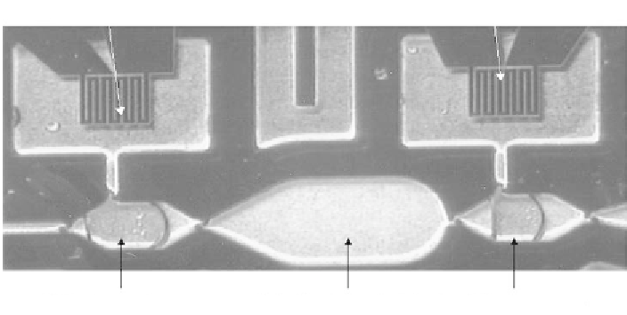
Figure 5.15
Enlarged view of the detection chamber and diffusion blocking reservoirs (courtesy of
LETI/BioMérieux).
locations in a capillary tube [7]. In this example of the simultaneous PCRs, the bio-
logical sample to be analyzed is brought into the tube under the action of capillary
forces (or by pipetting). At the solid wall, at different locations, different primers
(reverse and forward) have been grafted (Figure 5.16).
When the liquid has filled the tube and is at rest, the primers are released by
optical methods (insolation) and then diffuse locally inside the tube (Figure 5.17).
The presence of both reverse and forward primers is necessary for the PCR ampli-
fication. In the regions of the tube where a sequence of the DNA contained in the
liquid is corresponding to a type of primer, DNA amplification occurs and detection
is made by fluorescence methods.
Within the time frame of the PCR cycles for amplification, the primers should
not diffuse to the next region in a substantial way (Figure 5.18). If they do, detection
would be inaccurate. Thus, it may be necessary to introduce neutral gaps between
the primers regions so that there is no diffusion of primers between two regions.
However, the aim is a compact microsystem and these gaps should be reduced as
much as possible. The problem consists of calculating the primers diffusion inside
the tube and to determining their concentration as a function of time.
Figure 5.16
Schematic view of the capillary with the different labeled regions.