Environmental Engineering Reference
In-Depth Information
A
B
100
m
m
100
m
m
C
D
100
m
m
1 mm
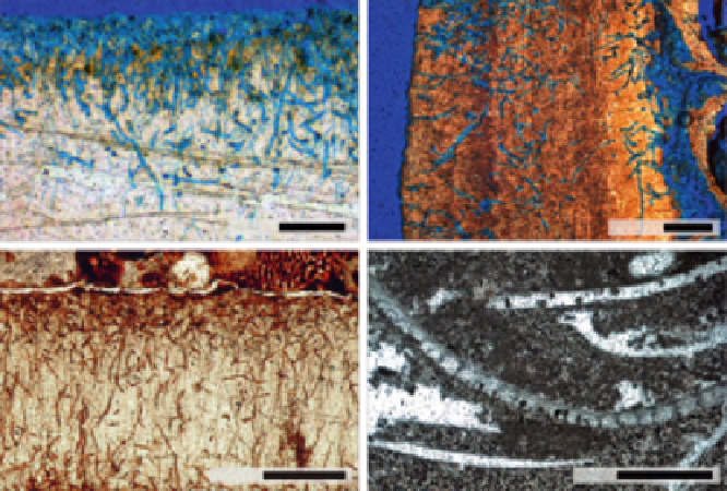
FIGURE 1
Microbioerosion in thin sections. (A) The ichnotaxa
Eurygonum nodosum
and
Scolecia
filosa
in a recent bivalve shell from the Azores. The shell was vacuum-impregnated with blue-
stained epoxy resin prior to sectioning. (B) The fungal trace
Orthogonum lineare
in a Pleistocene
cold-water coral from Rhodes, Greece. The microborings were still empty and accessible for stained
vacuum impregnation. (C) Same assemblage as in A, but from the Carboniferous Buckhorn Asphalt
Lagerst¨tte, USA. The microborings are naturally impregnated with asphalt. (D) The fungal micro-
boring
Saccomorpha clava
, well preserved in a brachiopod shell (bottom), and a completely recrys-
tallized bivalve shell (top) from the Silurian of Gotland, Sweden (courtesy of Axel Munnecke).
the sample in a vacuum chamber at about 10 kPa for a couple of minutes and
impregnation of the sample surface after cutting.
2.2 Vacuum Cast-Embedding
The most approved method for visualizing the 3D architecture of tunnel systems
left by microbioeroders is the vacuum cast-embedding technique (e.g.,
Nielsen
and Maiboe, 2000; Wisshak, 2006
), which produces polymer resin casts
(
Fig. 2
). These casts can be studied under the SEM in order to display even
the most delicate morphological features. A prerequisite for this technique is,
however, that the borings are still empty or filled only with little-lithified
matrix. Immersion in a tenside for several hours (e.g., Goldschmidt Rewo
GmbH: Rewoquat W3690), directly followed by a treatment with hydrogen per-
oxide, shatters and removes clayey matrix within the borings. Final cleaning in
an ultrasonic bath, rinsing in distilled water, and drying complete the preembed-
ment procedure. The samples are then placed in a small vacuum chamber
specifically designed for cast-embedding (Struers CitoVac or its precursor
Epovac). The chamber allows the infiltration of several samples with a


Search WWH ::

Custom Search