Image Processing Reference
In-Depth Information
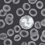
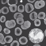
(a)
(b)
FIGURE 6.7
(a) Normal leucocyte images and (b) segmented leucocyte images using Type II fuzzy set.
(From
Micron
, 61, Chaira, T., Accurate segmentation of leukocyte in blood cell images using
Atanassov's intuitionistic fuzzy and interval Type II fuzzy set theory, 1-4, Copyright 2014,
with permission from Elsevier.)
Obtaining a ground truth image is very tedious and not perfect, but still it
gives a useful indication. For all the methods, the misclassification error is
calculated, which is defined as [25]
Error
=−
∩+∩
+
1
BBFF
BF
ET
GT
ET
GT
GT
GT
where
F
GT
and
B
GT
denote the foreground and background area pixels of the
ground truth image, respectively
F
ET
and
B
ET
are the foreground and background area pixels of the experi-
mental thresholded image, respectively
This error reflects the percentage of wrongly assigned pixels that ranges
from zero or no error, that is, when the image is exactly segmented to 1 or
when the image is wrongly segmented. The total area of the object pixels,
which are the cells shown in black, is obtained from the histogram of the
segmented image, and likewise, the background (white) pixels are com-
puted from the histogram of the segmented image. The unit of area is in
pixels. If the area covered by each pixel is known, that is,
x
(mm
2
/pixel),
then it is possible to convert to a physical unit. Ideally, the misclassification
error is zero.


Search WWH ::

Custom Search