Biomedical Engineering Reference
In-Depth Information
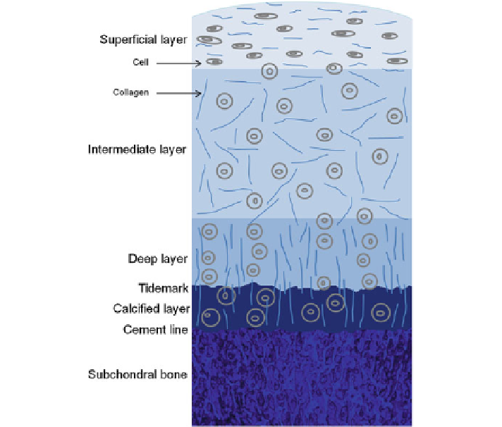
Fig. 2.3
Illustration of the structure of an OC tissue showing the arrangement of the cells and
collagen fibres within the articular cartilage. Articular cartilage has four layers: the superficial
layer, the intermediate layer, the deep layer, and the calcified layer. The superficial layer is the
thinnest layer with flattened chondrocytes and collagen fibres that are parallel to the surface. The
intermediate layer represents the thickest layer of the cartilage with spherical chondrocytes and
thicker collagen fibrils, which are randomly aligned. The deep layer has spherical chondrocytes
that are aligned in columns and collagen fibrils are parallel to each other and perpendicular to the
articulating surface. The calcified layer is a thin layer with hypertrophic cells and separated from
the deep layer by the tidemark. Subchondral bone is located below the cartilage while the cement
line forms the interface between the calcified layer and the subchondral bone
2.3 Bone and Subchondral Bone
Bone
is composed of organic and inorganic components. Minerals, principally hy-
droxyapatite (HAp), comprise 50-70% of the bone, collagen type I rich organic
matrix 20-40%, water 5-10%, and lipids less than 3% [
25
]. HAp contributes to the
rigidity and load-bearing strength, while the organic matrix provides flexibility and
elasticity of the tissue. Four cell types are present in bone: osteoblasts, osteoclasts,
osteocytes and bone lining cells. Osteoblasts are the mature bone-forming cells found
on the bone surface, while osteocytes are embedded in the lacunae encircled with