Biomedical Engineering Reference
In-Depth Information
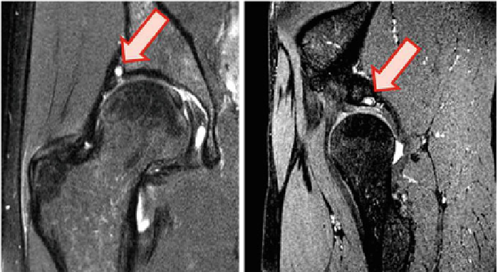
Fig. 10.8
Radiological analysis: Diagnosis of acetabular and labral lesions (
arrow
) in the posterior
part of the acetabular rim
the femoral head and
O
starts to exceed the radius (
r
) of the femoral head. Deviation
from the normal geometry is usually associated with larger
60
∈
).
Based on subject-specific data (MRI and 3D bones reconstruction), these standard
measurement methods were numerically implemented, improving the (subjective)
reading of medical images. The dancer hip was thus analyzed, according to those
two anatomical parameters. No morphological abnormalities were detected and it
was concluded that the measured hips have an average positive depth (left hip: 8.16
mm, right hip: 7.89 mm) and an average
α
angles (
>
15
∈
<α<
α
angle in the normal range (36
.
43
∈
). The results were validated by radiological experts.
The same radiological experts performed consensus readings of the subjects' MR
images [
5
]. The acetabular cartilage and labral abnormalities were assessed qualita-
tively. For this subject, acetabular and labral lesions were diagnosed in the posterior
part of the acetabular rim (see Fig.
10.8
). To describe the exact location of the lesions,
the acetabulum was divided into eight sectors (1: anterior, 2: anterosuperior, 3: supe-
rior, 4: posterosuperior, 5: posterior, 6: inferoposterior, 7: inferior, 8: anteroinferior),
as depicted in Fig.
10.7
.
54
.
10.4 Biomechanical Analysis of Professional Ballet Dancer
Hip Joint
This study was conducted in collaboration with doctors from the department of Radi-
ology and department of Orthopedic Surgery of the University Hospital of Geneva
and female professional ballet dancer from the ballet of the Great Theater of Geneva.
The developed approach is used to analyze the mechanical behavior of articu-
lar layers of a dancer's hip joint. The choice of subject is justified by the nature