Biomedical Engineering Reference
In-Depth Information
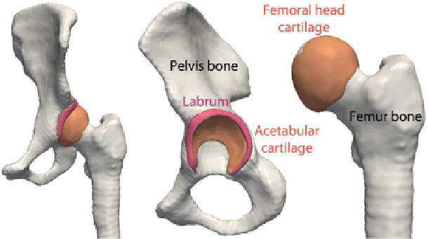
Fig. 10.1
Hip joint anatomy: bones and cartilage layers
To understand the human joints mechanism and thoroughly investigate the devel-
opment of OA, several studies were conducted. Different disciplines (e.g., medi-
cine, biology, biomechanics and applied sciences) are involved in the exchange and
combination of knowledge from different expertise domains. Despite the growing
advancements, limitations still exist and much work remains to be done to better
respond to the complexity of both the human anatomy and medical procedures.
10.1.1 Medical Context
This study focuses on the hip joint, which is crucially important in the musculoskele-
tal system. The hip joint supports the weight of the body in both static and dynamic
postures. It allows for a large range of movement and for the transfer of high forces
between the femur and the pelvis during daily activities [
3
]. The hip joint is clas-
sified as a ball and socket joint, with the acetabulum acting as the socket in which
the spherical femur head articulates (see Fig.
10.1
). Both bone surfaces are covered
with an articular cartilage which prevents direct bone-to-bone contact and allows a
pressure distribution inside the joint. Connected to the acetabulum rim, the acetabular
labrum is a fibrocartilaginous structure that increases the acetabulum depth, grips the
femoral head and provides stability to the hip joint. The hip joint is further reinforced
by ligaments [
4
]. Given its role in the MS, the hip joint is especially vulnerable to
different pathologies and mostly OA. Although the frequency of hip OA increases
with age, OA is not exclusively an aging process as it is also seen in younger patients
[
5
]. In fact, the damage of the labrum or labral tear was associated with the develop-
ment of hip OA. Studies have shown that a labral tear is frequently found in younger
patients, while for older patients the labral tear is more often associated with chondral