Environmental Engineering Reference
In-Depth Information
coating acts as a protective layer to avoid direct contact of the magnetite core
with the liquid to be treated. The negative charge of the silica-coated surface, on
the other hand, facilitates the assembly of cation surfactant micelles arisen from
electrostatic attraction. After molecular templating, the zeta potentials of the
resultant particles were positive over a wide pH range. This resembles the zeta-
potential variation of an oil droplet or air bubble with pH in CTAC aqueous
solutions. After sol-gel reaction to fill the voids observed in the AFM images
(Fig. 6.9b), the particles become negatively charged again. The measured IEP of
the collected particles is at pH 4.0, which is different from that for a silica
surface. This observation suggests that the trapped CTAC template partially
balances the negative charge of silica in the templates voids. After calcination,
the surfactant was removed, leaving behind the silica network on the magnetite
surface, which has the IEP at pH 2.6, identical to the IEP value for silica. This
finding confirms a full coverage of magnetite by silica, making the coated
surface silica-like. To further confirm the protection of magnetite particles by
the two-step silica coatings, the coated particles were immersed in a 1 M acid
solution. The results showed a negligible amount of iron being leached out after
a 12 h immersion, indicating good protection of magnetite by silica coatings.
To further verify the necessity and success of each step described in Fig. 6.7,
the samples collected at various stages of the synthesis are characterized by
DRIFT and XPS. The results can be found in our related publications [122].
The morphology of Fe
3
O
4
and mesoporous silica-coated Fe
3
O
4
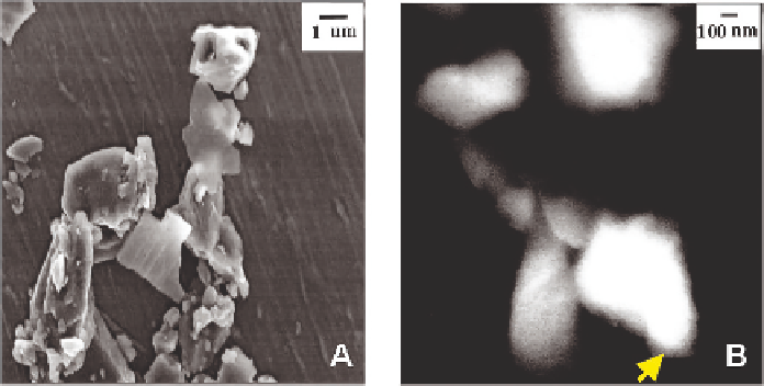
is observed
by TEM. Figure 6.11 shows that bare Fe
3
O
4
has a well-defined crystalline
feature with sharp edges and corners. In contrast, the TEM image of mesopor-
ous silica-coated Fe
3
O
4
shows diffuse edges. Combined with the evidence from
Fig. 6.11 TEM images of untreated Fe
3
O
4
particles (A) and Fe
3
O
4
particles with template-
assisted silica coatings (B). Micrograph (B) is a dark-field image, obtained with tilt
illumination