Biology Reference
In-Depth Information
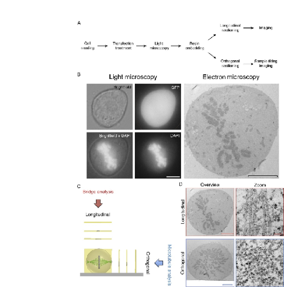
FIGURE 20.1
CLEM performed on mitotic cells. (A) A workflow to achieve CLEM using longitudinal or
orthogonal sectioning. (B) A transfected mitotic HeLa observed by LM (Brightfield, GFP, and
DAPI) and by electron microscopy. Scale bar 5 mm. (C) Schematic of longitudinal and
orthogonal EM sectioning, and examples of output analysis. (D) Representative electron
micrographs of cells sectioned longitudinally (above) and orthogonally (below) with high
magnification of microtubules (right). Scale bar 4
m
m (overview) and 50 nm (zoom).