Biology Reference
In-Depth Information
Covalent binding in cells can also be examined using flow cytometery. This is done
by monitoring cells after MSA treatment and comparing the number of cells in
the G
2
/M phase of the cell cycle after drug treatment and after drug washout. If the
MSA is covalently bound, then the cells will remain in G
2
/M even after washout of
the unbound drug. This method is described in
Buey et al. (2007)
(
Fig. 19.6
).
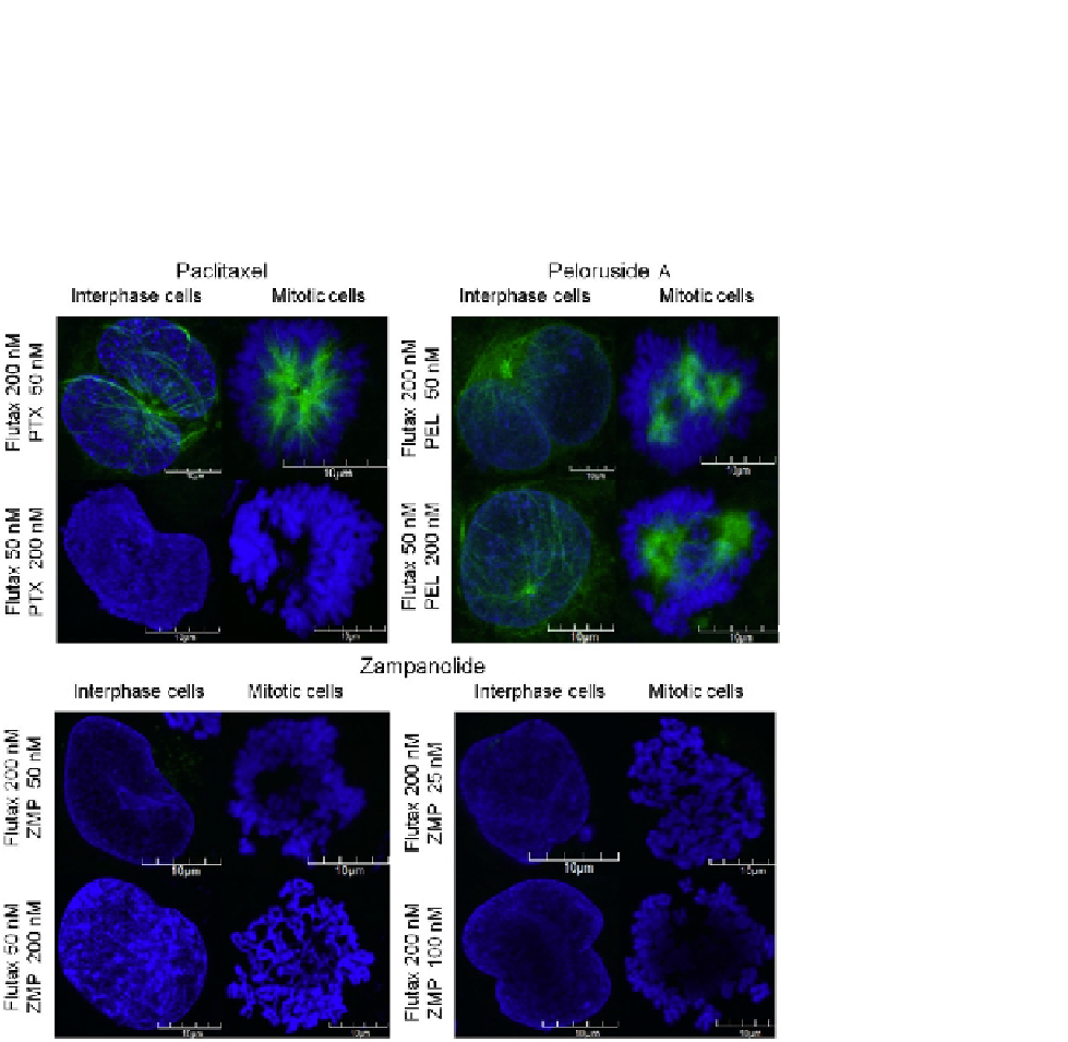
FIGURE 19.6
Flutax-2 staining of HL-60 cells showing covalent binding of ZMP. Flutax-2 staining is shown
in light gray (or green for color images). DAPI staining of the nucleus is in dark gray (or blue).
When in excess, Flutax-2 stains the MTs in both paclitaxel (PTX)- and peloruside (PEL)-
treated cells. When paclitaxel is in excess, Flutax cannot stain the MTs due to competition for
the taxoid binding site; however, when peloruside is in excess, Flutax-2 can still stain the MT
network as both ligands can bind simultaneously. Lack of MT staining by Flutax-2 in
ZMP-treated cells indicates the irreversible, covalent binding of ZMP, as even in excess,
Flutax-2 cannot compete with ZMP to stain the MTs.