Biology Reference
In-Depth Information
The persistence length calculated following Eq.
(2.1)
is related to the Young's
modulus,
E
, of the microtubule by
l
p
¼
EI
2
(2.3)
kT
where
I
is the moment of inertia of the microtubule and
kT
is the thermal energy of
the solution. The goal, then, is to measure
y
s
along an individual microtubule and use
Eq.
(2.1)
to calculate the persistence length.
However, in solution, microtubules fluctuate rapidly, making high-precision
measurements of
y
s
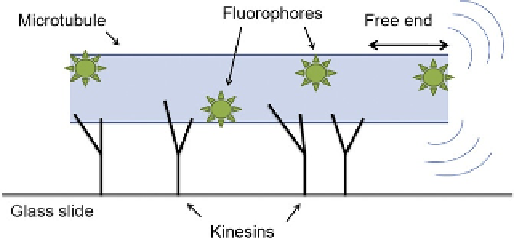
difficult. Our gliding assay uses the motor protein kinesin-1
to propel a microtubule over a glass surface (
Fig. 2.2
). If the density of kinesin
is high enough, the microtubule will be bound, on average, to many kinesins.
However, the microtubule tip will fluctuate before attaching to the next kinesin
the tip comes in contact with. The tip fluctuations are well described by
Eq.
(2.1)
, and the rest of the microtubule simply follows the same path as the
tip since kinesin is processive (remains bound to the microtubule for many
ATP turnovers). Thus, the trajectory of the microtubule is a frozen-in fluctuation
of the tip—the persistence length of the trajectory reports the persistence length of
the tip (
Duke, Holy, & Leibler, 1995
). Gliding assays have been used previously to
calculate microtubule persistence lengths (
van den Heuvel et al., 2007
); this
method uses a different (and, arguably, simpler) method of reconstructing the
microtubule trajectory.
In order to reconstruct the microtubule trajectory, we first attach single fluoro-
phores sparsely to the microtubule surface. Using a microscope capable of imaging
single fluorophores (a total internal reflection fluorescence, TIRF, microscope in our
case), we track all individual fluorophores on a microtubule (
Crocker & Grier, 1996
)
FIGURE 2.2
Cartoon of kinesin gliding assay for microtubules. Kinesins are specifically attached to a glass
slide by the coiled-coil, leaving the motor domains free to contact microtubules. Microtubules
are propelled by kinesins; the free microtubule end fluctuates due to thermal fluctuations.
Single fluorophores are sparsely attached to the microtubule; individual fluorophores are
imaged using a TIRF microscope.