Biology Reference
In-Depth Information
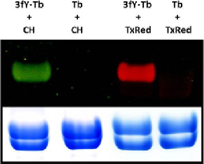
FIGURE 1.2
Detection of fluorophore-labeled tubulin: 3-Formyltyrosinated tubulin (3fY-Tb) or unmodified
tubulin (Tb) in PME buffer was incubated with CH or TxRed in the presence of 10 mM
4aFasdescribedprior toSDS-PAGEanalysis. Thegel was thenvisualizedunder long-wavelength
UV light (top) and stained with Coomassie blue (bottom). The mass of protein loaded into each
well was 25
m
g. Photographic exposure time for the fluorescent gel
¼
10 s.
FIGURE 1.3
Detection of biotinylated tubulin: 3-Formyltyrosinated tubulin (3fY-Tb) or unmodified tubulin
(Tb) was incubated with biotin hydrazide (BH) in the presence of 4aF as described prior
to SDS-PAGE analysis. Top: Western blot with streptavidin-HRP conjugate (Life
Technologies). Bottom: Coomassie stain. The mass of protein loaded into each well was 1
m
g
for the Western blot and 5
m
g for the protein stain.