Biology Reference
In-Depth Information
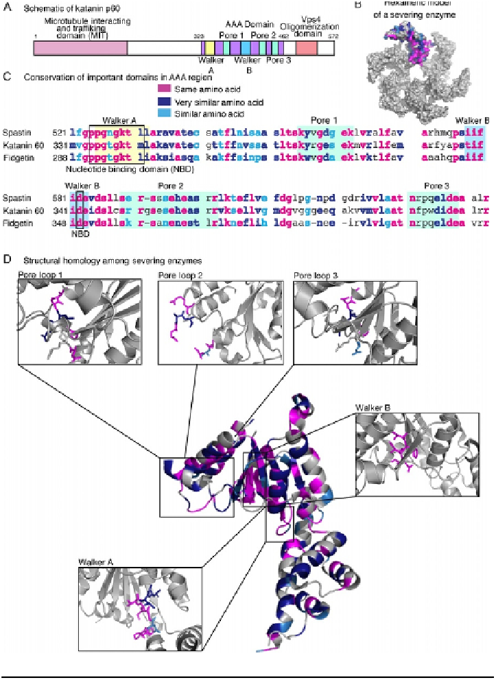
FIGURE 13.1
Conservation of domains in all known severing enzymes. (A) Schematic of katanin p60
showing the microtubule interacting and trafficking (MIT) domain (pink), ATPases associated
with diverse cellular activities (AAA) domain (purple) with the Walker A domain (yellow) and
Walker B domain (blue), the regions associated with the pore loops (green), and the Vps4
oligomerization domain (orange). Spastin and katanin have similar domain structure,
although it is important to note that fidgetin's MIT domain has not been identified yet. (B)
A model of the hexameric structure that severing enzymes are thought to adopt.
Conserved residues (pink), very similar residues (dark blue), and similar residues (blue)
are denoted in the shading comparing Drosophila melanogaster proteins. Note that this