Biology Reference
In-Depth Information
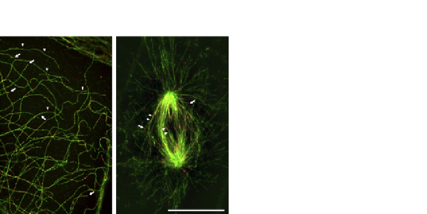
FIGURE 10.1
GTP-tubulin labeled with MB11 antibody (red) on PTK2 cells stably expressing GFP-tubulin
(green) imaged on a structured illumination microscope (SIM). Acquisitions were performed
in 3D SIM mode, with an n-SIM Nikon microscope before image reconstruction using the
NIS-Elements software based on
Gustafsson et al. (2008)
. The system is equipped with
an APO TIRF 1001.49 NA oil immersion and an EMCCD DU-897 Andor camera. Arrows
point at GTP caps and arrowheads show GTP-tubulin islands, both stained with the MB11
antibody. Signals have been artificially increased for better visualization in the black and white
version of the figure.
using a nonionic detergent in an effective microtubule-stabilizing buffer. Using only
a PIPES-based buffer is not sufficient to preserve a large proportion of cellular mi-
crotubules for more than a few minutes, thus glycerol and optionally Taxol are also
added throughout the labeling operations (
Fig. 10.1
). To allow multiple labeling, cell
fixation can be achieved as soon as incubation with MB11 and consecutive rinses
have been performed. GTP-tubulin staining with MB11 is also compatible with
the expression of GFP-tubulin (
Fig. 10.1
).
10.1.1
Permeabilization
Cells are grown in culture medium at 37
C, 5% CO
2
. Replace the culture
medium by warm permeabilization buffer. Incubate exactly 3 min at 37
C. Wash
twice 1 s with caution in PEM-G, holding the coverslip.
Optionally, 1
m
M Taxol can be added to the extraction, incubation, and washing
solutions, especially if labeling is to be correlated with prior
in vivo
imaging.
Note that, in contrast with what happens when Taxol is added during microtubule
assembly or when using it at high concentration (
Dimitrov et al., 2008
), low doses
of Taxol do not alter the number and length of GTP-tubulin islands
a posteriori
.
Put the coverslip directly on the drop of MB11 antibody.