Biology Reference
In-Depth Information
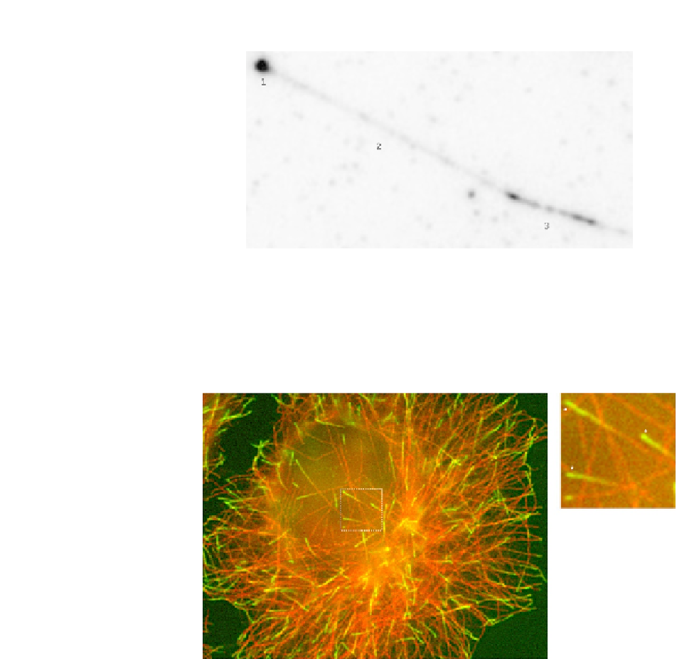
FIGURE 8.6
An in vitro experiment with 75 nM 6
HIS-GFP-EB3 as imaged via TIRF microscopy. The EB
comet is clearly visible at the plus end (1), as are lattice interactions along the length of the
growing MT (2). The GMPCCP-stabilized seed is visible in (3), where the EB tends to bind
more strongly than the GDP lattice, presumably due to the GMPCCP moiety. The imaging
conditions were 3 mW 491 nm laser, 100
objective, 500 ms exposure time, 37
C.
FIGURE 8.7
GFP-EB1 expressed in HeLa cells and imaged via TIRF microscopy. Cells were fixed with
methanol and 1 mM EGTA and stained with anti-EB1 (green) and antityrosinated-
-tubulin
a
(red). The EB comet is visible at the plus end (*).
-casein blocking or an issue with either tracer tubulin or the seeds (e.g., lack of cen-
trifugation to a pellet prior to loading, reuse of diluted seeds). As a comparison to the
in vitro
assay,
Fig. 8.7
shows EB1 comets produced in a HeLa cell. Again the EB
comets are clearly visible and most importantly they are comparable to those produced
with the
in vitro
assay.
k