Biology Reference
In-Depth Information
7.2
MANUFACTURING OF MICROPATTERNED COVERSLIPS
Work in a dedicated clean area, preferably under a hood. Wear gloves and avoid dust
by covering the photomask and coverslips. We prefer square coverslips because they
are easier to orient; however, round coverslips will also work well.
Figure 7.1
A
summarizes how the micropatterns on photomask is transferred to the coverslip.
A
Deep UV
Quartz photomask
ddH
2
0
PEG-PLL
Glass coverslip
B
Adhered cell
Fibronectin
Carboxyl surface
Glass coverslip
C
Fibrinogen-Cy5
Phase contrast
10
m
m
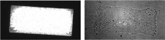
FIGURE 7.1
Deep UV micropatterning technique standardizes cell shape and size for subsequent imaging
of mitochondria and microtubules. (A) Schematic of the deep UV technique to transfer the
micropatterns from the photomask to the PEG-PLL-coated glass coverslips. The UV
irradiation creates carboxyl groups in the exposed areas of the PEG, which subsequently
binds to proteins such as fibronectin. (B) Schematic of UV-exposed PEG-PLL-coated
coverslip treated with fibronectin then seeded with cells. The fibronectin binds only to
the carboxyl surface of the PEG-PLL. Cells adhere only to the fibronectin. (C)
Phase-contrast image of an RPE1 cell adhering to the rectangular shape coated with
fibronectin. Fibrinogen-Cy5 was added to the fibronectin solution to help visualize the
shape of the coated surface.