Biomedical Engineering Reference
In-Depth Information
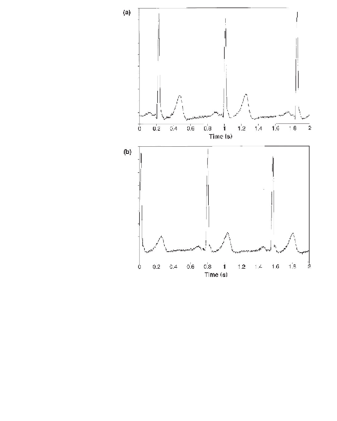
Figure 1.14
Single-lead ECG recordings: (a) using an Ag/AgCl standard bioelectrode; (b) using
the capacitive active bioelectrode. (Reprinted from Prutchi and Sagi-Dolev [1993], with permission
from the Aerospace Medical Association.)
is built around one-half of two TL064 quad op-amps. Eight copies of this circuit
constitute the 16 identical biopotential ampli
cation channels. Operation of a single
channel is described in the following discussion.
A biopotential signal detected by a bioelectrode is coupled to the noninverting inputs of
fi
the
er. The input impedance is given
mostly by the input impedance of the front-stage op-amps, yielding
fi
first-stage ampli
fi
er and the shield driver ampli
fi
100 M
Ω
paralleled
with 100 pF. R1 limits the current that can
flow through the input lead, while diodes D1
and D2 shunt to ground any signal that exceeds their zener voltage. This arrangement pro-
tects the inputs of the ampli
fl
fi
ers from ESD and from the high voltages present during car-
diac de
brillation. Furthermore, it protects the subject from currents that may leak back
from the ampli
fi
ers or associated circuitry.
The shield driver is con
fi
er. The actual drive, however, deter-
mined by R2 and R3, is set to 99% of the signal magnitude at the inner wire to stabilize
fi
gured as a unity-gain bu
ff

Search WWH ::

Custom Search