Biomedical Engineering Reference
In-Depth Information
where
n
is the refractive index of the medium in which the lens is working and
a
is the half-angle of the
maximum cone of light that can enter or exit the lens.
The magnification of the microscope system depicted in
Fig. 8.2
is the product of the magnifi-
cations of the objective lens 1 and the eye piece lens 2:
LL
total
f
1
f
2
M
¼
(8.8)
where
L
is the distance between the second focal point of the objective lens (lens 1) and the first focal
point of lens 2 (also called the tube length),
L
total
is the near-point distance (also called the viewing
distance), and
f
1
and
f
2
are the focal lengths of the lens 1 and lens 2, respectively. The brightness of the
recorded image is inversely proportional to the square of the magnification:
1
M
2
:
If
(8.9)
According to
(8.7)
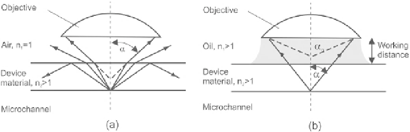
, the numerical aperture of the objective lens depends on the refractive index of the
coupling medium between the lens and the device and cannot be more than unity. In order to increase
the numerical aperture, oil immersion objective lenses are often used.
Figure. 8.3
shows that the
refractive index of the oil and the substrate material of the device increase the semi-angle of the cone
of rays from the point source leading to a higher effective NA. While the maximum achievable
numerical aperture in air is about 0.85, immersionobjectivelensesmayreachamaximumnumerical
aperture of 1.4. Oil immersion objective lenses are designed to work with a given oil as a working
medium:
a
c
¼
arcsin
ð
n
1
=
n
2
Þ:
(8.10)
The effective field of view of a measurement based on a microscope/camera system depends on both
the objective lens and the surface area of the sensor. For a given magnification
M
and sensor surface
area
A
s
, the effective area of view is
M
2
A
v
¼
A
s
=
:
(8.11)
FIGURE 8.3
Typical situation of a micromixer with a transparent substrate material: (a) in air and (b) immersed in oil.


Search WWH ::

Custom Search