Biomedical Engineering Reference
In-Depth Information
4
Micropatterned Supports to Control Cell Adhesion
4.1
Engineering Cell Shape and Adhesive Area
Micropatterning techniques have been extensively applied to engineer cell
position, shape, and adhesive area [89, 90]. These approaches generally rely
on creating domains that readily adsorb proteins, and hence are cell-adhesive,
and are surrounded by a nonfouling, nonadhesive background. These mi-
cropatterned supports can be easily generated by conventional photolitho-
graphy as well as “soft” lithography approaches, including microcontact
printing. In addition, direct protein stamping has been applied to create
cell adhesive domains, but the stability of these patterns is limited by cell-
mediated ECM reorganization and deposition. These substrates with defined
adhesive areas have been exploited to analyze the roles of cell shape and cell-
cell interactions on cell survival, expression of tissue-specific markers, and
commitment to differentiated lineages [91-94]. Conversely, micropatterned
substrata allow engineering of adhesive area, and in particular focal adhe-
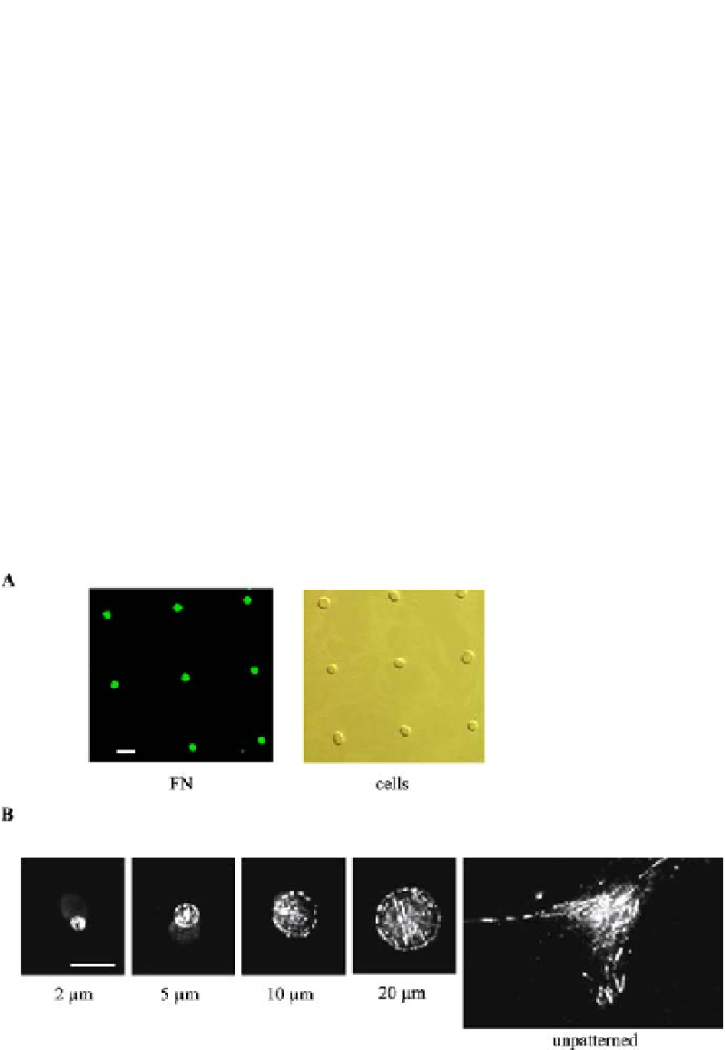
Fig. 7
Micropatterned surfaces to engineer focal adhesion size.
A
Adhesive islands within
nonfouling background showing preferential FN adsorption and cell adhesion. Cells
adhere and remain constrained to micropatterned island.
Bar
: 20
m.
B
Vinculin local-
ization to micropatterned domain, showing precise control over focal adhesion assembly.
Bar
: 10
µ
µ
m. Adapted from [95, 99]