Biomedical Engineering Reference
In-Depth Information
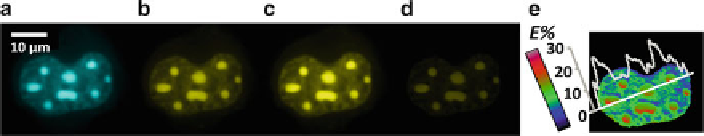
Fig. 3.3
Demonstration of the homodimerization of the CCAAT/enhancer-binding protein alpha
.C=EBP'/ in live mouse pituitary cell nuclei using widefield FRET microscopy. Images from the
nucleus of a cell coexpressing CFP-C/EBP˛ (FRET donor) and YFP-C/EBP' (FRET acceptor)
were acquired in the donor (a), acceptor (b), and FRET (c) imaging channels using an Olympus
IX70 widefield epifluorescence microscope equipped with a
60/1.2NA water objective lens, a
ldgi-xcite.com
)
. The processed FRET (PFRET, d) and apparent FRET efficiency (E%, e) images
were obtained after processing the data with the PFRET algorithm in combination of the images
acquired from the single-label expressing cells (see Sect.
3.3.4
). The interaction between CFP-
and YFP-tagged C/EBP˛ is demonstrated by the E% image, indicating the homodimerization of
C/EBP˛ in regions of centromeric heterochromatin of cell nucleus. For each imaging channel,
the same dichroic mirror designed for imaging both CFP (445-485 nm) and YFP (520-580 nm)
was used, and the excitation and emission filters were 436/20 nm and 470/30 nm (donor channel),
500/20 nm and 535/30 nm (acceptor channel), and 436/20 nm and 535/30 nm (FRET channel). See
Sect.
3.4.6
for more details about the C/EBP' biological model (Adapted from [
43
])
CAT scan) from a thick specimen [
48
,
49
]. For example, a laser scanning confocal
microscope (LSCM) focuses a laser beam on a single point of a specimen through an
objective lens and uses a point detector - usually a photomultiplier tube (PMT) and
occasionally an avalanche photodiode (APD); a pinhole is placed before the detector
to reject the out-of-focus light (light signal above and below the focal plane); a 2-D
image is obtained by moving the laser beam over a region of interest of the specimen
through the XY raster point-scanning mechanism; a stack of 2-D images at the
different depths of the specimen are acquired by moving the microscope objective
lens or stage along the optical axis. Compared to LSCM, a spinning-disk confocal
microscope can provide a much faster imaging speed since it employs a number
of pinholes designed in a specific pattern to focus laser beams at multiple points
of a specimen and uses a charge-coupled device (CCD) camera to acquire a 2-D
image [
49
-
51
]. Confocal microscopy has been applied to a wide range of biological
applications [
46
] - an example of applying an LSCM to study the receptor-ligand
binding and internalization based on FRET [
52
-
54
] is shown in Fig.
3.4
(see
Sect.
3.3.4
). Many aspects related to confocal microscopy imaging techniques and
data analysis for various applications are described in the literature [
55
].
3.3.3
Two-Photon Excitation (TPE) Microscopy
Two-photon absorption was theoretically predicted by Goppert-Mayer in 1931.
TPE imaging experiments in a laser scanning confocal microscope were first
demonstrated in 1990 [
56
]. In a TPE event, two photons, each of which carries
Search WWH ::

Custom Search