Biomedical Engineering Reference
In-Depth Information
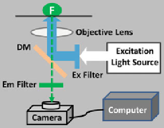
Fig. 3.2
The basic setup of an inverted widefield epifluorescence microscope. In widefield
epifluorescence microscopy, imaging a fluorescent sample (F) requires a correct combination of the
excitation (Ex) and emission (Em) filters and a dichroic mirror (DM), which is chosen based on the
Ex and Em spectra of the fluorophore. The specific Ex wavelengths of light for the fluorophore are
selected from the Ex light source (such as an arc lamp) passing through the Ex filter; the selected
light is then reflected by the DM to excite the F through the objective lens (blue line trajectory);
the light emitted from the excited fluorophores and collected by the same objective (
green dashed
line
) will transmit through the DM and the Em filter before reaching the detector (usually a charge-
coupled device (CCD) camera)
events in living cells using fluorescent reporters as long as the axial resolution is
not a concern. For example, imaging genetically encoded fluorescent indicators
(chameleons) for Ca
2C
using widefield microscopy has allowed the measurements
of Ca
2C
signals in the cytosol and organelles [
36
-
38
]. An example of applying
widefield microscopy to study protein-protein interactions in living cells based
on FRET is shown in Fig.
3.3
(see Sect.
3.3.5
). The axial resolution in widefield
microscopy is poor due to the out-of-focus light contamination and can be improved
by applying iterative constrained 3-D deconvolution techniques [
39
-
42
].
3.3.2
Single-Photon Excitation (SPE) Confocal Microscopy
The principle of SPE confocal microscopy or laser scanning confocal microscopy
(LSCM) imaging was introduced in 1957 by Marvin Minsky. The invention of
laser in the 1960s and advances in laser technologies in the past 50 years have
made confocal microscopy a major imaging tool for biologists today [
44
-
46
]. For
example, a new supercontinuum laser (also called white-light laser) tunable from
470 to 670 nm in 1 nm increments has been equipped in Leica TCS SP5 X confocal
microscopes, giving scientists more choices of imaging with various fluorescent
probes compared to a regular confocal microscope that only carries fixed laser lines
[
47
]. Compared to conventional widefield microscopy, confocal microscopy offers
much higher axial resolutions by using spatial filters to eliminate or minimize the
out-of-focus light and provides the capability to collect serial optical sections (like
Search WWH ::

Custom Search