Biomedical Engineering Reference
In-Depth Information
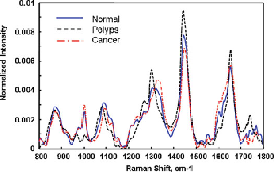
Fig. 1.22
Mean NIR Raman spectra from normal (n D 324), polyps (n D 184), and adenocarci-
noma (n D 309) colonic tissues, respectively (Adapted from Widjaja et al. [
65
], with permission)
1.4.4
Oral Cancer Diagnosis
Raman spectroscopy has been used for diagnosis of oral mucosa diseases. The
Raman spectra of
in vivo
oral mucosa were investigated by Schut et al. using a
mouse model [
68
]. In their experiment, dysplasia was first induced into the mouse
palate epithelium, and then the Raman spectra were assessed at various dysplastic
stages. The spectra of the mouse oral mucosa were obtained from 10 to 100 s.
They found a specificity of 93% and a sensitivity of 78% for detecting low-grade
dysplasia and a specificity of 100% and a sensitivity of 100% for detecting high-
grade dysplasia/carcinoma
in situ
. Malini et al. evaluated the ability of Raman
spectroscopy to differentiate normal, inflammatory, premalignant, and malignant
oral lesions of biopsied tissue [
69
]. A set of the Raman spectra was shown in
Fig.
1.23
. Each spectrum was obtained with an integration time of 600 s. Data
analysis of these spectra showed that the ability to distinguish among pathologic
conditions was poor. In another independent study of
ex vivo
human mucosa samples
by de Veld et al. [
70
], similar conclusion was obtained. Very recently, Guze et al.
[
71
] studied the Raman spectra of human normal oral mucosa among anatomic oral
sites and among subjects of different races and gender. High-quality Raman spectra
at the high-frequency range could be obtained within 1 second using an
in vivo
fiber-based Raman probe similar to the endoscopic lung Raman probe (Fig.
1.13
).
The averaged Raman spectra of oral cavity of different anatomic sites are shown in
Fig.
1.24
. They found that
in vivo
Raman spectra taken from the oral cavity did not
show strong differences between Asian and Caucasian subgroups, but the spectra for
different oral sites within the same ethnic group were different and separable. Their
results demonstrated that the lack of sensitivity in the previous
ex vivo
study was due
to the variability in the spectra produced by the different tissue types of the mouth
and oropharynx. Further study is warranted for the potential of Raman spectroscopy
for
in vivo
oral cancer diagnosis, including the low-frequency fingerprint region.
Search WWH ::

Custom Search