Biomedical Engineering Reference
In-Depth Information
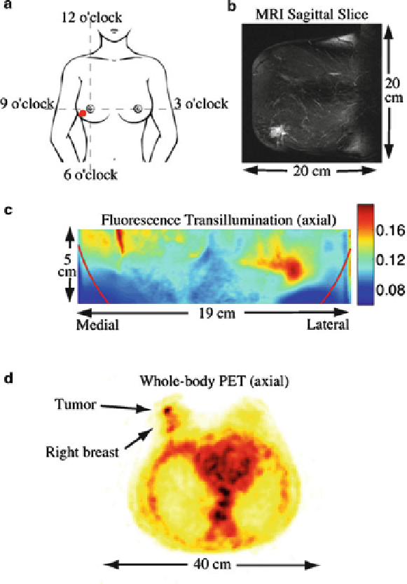
Fig. 10.6
(
a
) Illustration of the tumor location. (
b
) Gadolinium-enhanced sagittal MR image slice
showing the tumor in the lower
left corner
.(
c
) Fluorescent transillumination image (explained in
text). (
d
) Axial slice from 18F-FDG whole body PET image. The view is from the patient's feet
(i.e., the right breast appears on the
left side
) (Figures (
a
-
c
) reprinted with permission from [
64
,
88
].
c
2007, Optical Society of America
low-resolution and partial volume effects. Only recently, the combination of optical
imaging with other clinical functional imaging modalities has been considered.
Especially, the combination of PET with DOI is currently being pursued. However,
to date, the combination of both techniques is done nonconcurrently and relies on
software registration.
Search WWH ::

Custom Search