Biomedical Engineering Reference
In-Depth Information
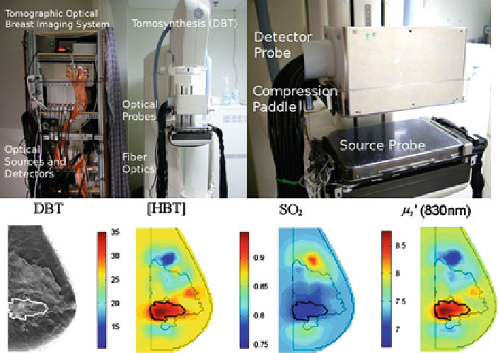
Fig. 10.4
Optical imager integrated with digital breast tomosynthesis. Example of DBT and
optical functional parameters Reconstructed image sections for the right breast in 45-year-old
woman. The breast contains a 2.5-cm invasive ductal carcinoma (highlighted by the thick black
margin).
probe that is mechanically scanned by the operator over the region of interest.
The small dimension and flexibility of the US probes allow for an easy optical-
US coupling without modifying the standard US operating procedures, though
optical interrogation of tissue is then performed in the reflectance geometry which
is not optimal to probe deep tissue and which is very sensitive to tissue/optode
coupling.
A typical implementation of DOI integrated with ultrasound is depicted in
Fig.
10.5
. This system developed at the University of Connecticut is based on a
commercial US transducer that is mounted on the center of a custom handheld probe
with the optodes distributed at the periphery. The light delivery and light collection
is performed via optical fibers to allow for a small footprint and easy cleaning.
The optodes are distributed over the surface of the probe to offer multiple distance
measurements required to image deep-seated tissue (a couple of centimeters).
The probe is holding 12 optical source fibers, 4 optical detector fibers, and 20
piezoelectric crystals comprising the US array. The optical and US systems can
operate simultaneously for coregistration without interference.
The structural information provided by the US imaging system is used to guide
the optical imaging reconstruction process. The lesion is localized on the US image,
and the lesion volume is segmented in a refined mesh whereas the nonlesion
Search WWH ::

Custom Search