Biomedical Engineering Reference
In-Depth Information
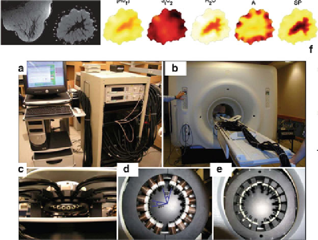
Fig. 10.3
The Dartmouth NIR/MRI systems. (
a
) Photograph of the portable NIR instrumentation
and control console. (
b
) Optical fibers extend from the system into the MRI. (
c
) Open architecture
breast array coil houses the optical fiber-positioning system. (
d
)Thefirst-and(
e
) second-
generation MR-compatible fiber-positioning mechanisms. (
f
) One set of breast MRI/NIR images
(Used with permission from [
75
])
between MR structural contrast and optical contrast (hemoglobin and water) has
been demonstrated [
73
] supporting the rationale of using MR-derived structural
priors in the optical inverse problem.
However, the current focus of MRI-optical fusion lies beyond the need to
improve optical imaging performances. Especially, the field of MR is very dynamic
with constant progress toward in vivo high-resolution functional imaging. There-
fore, present efforts from the optical imaging community focus on identifying
the complementary information that optical imaging can offer to MR imaging. In
the context of breast oncology, MRI is an efficient modality to detect lesions but
suffers from a lack of specificity (around 67 %) [
74
]. Conversely, optical imaging
techniques demonstrate promises in providing diagnostic functional information to
characterize the suspicious lesion. Stand-alone systems have been shown to provide
up to 88 % specificity [
75
], and the integration of multispectral optical imaging with
MRI may lead to increased optical specificity [
76
].
Similarly, functional MRI (fMRI) may benefit greatly from concurrent optical
acquisition. Especially, fMRI studies based on BOLD are limited at mapping
relative changes in tissue oxygen saturation, whereas optical imaging techniques
are able to quantitatively map both oxygenated and deoxygenated blood at high
frame rates (up to a few Hz). Therefore, optical imaging can provide complementary
Search WWH ::

Custom Search