Biomedical Engineering Reference
In-Depth Information
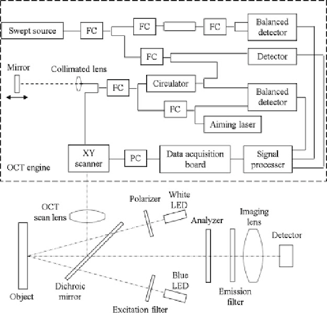
Fig. 9.17
Diagram of multimodal imaging system.
OCT
engine is inside the dashed box.
FC
fiber
coupler
9.4.7
Applications in Caries Detection
This multimodal imaging system has the advantage that the user can screen the
tooth or oral cavity with visible polarized light to obtain a visible reflectance image,
just as a conventional intraoral camera, or screen the tooth with blue excitation
light to view the fluorescence image. With polarized white light reflectance image
and fluorescence image and possibly with further image processing, the user can
identify healthy, carious, and suspicious regions quickly. Once the carious and
suspicious regions are located, OCT imaging can then be used to scan those regions.
For carious regions, OCT images can provide more detailed information, such as
decay depth, size, and boundary. For suspicious regions, OCT images can verify
whether the regions are indeed carious lesions, and if so, how deep the lesions are.
All three images, reflectance, fluorescence, and OCT, can be saved for progression
monitoring.
Search WWH ::

Custom Search