Biomedical Engineering Reference
In-Depth Information
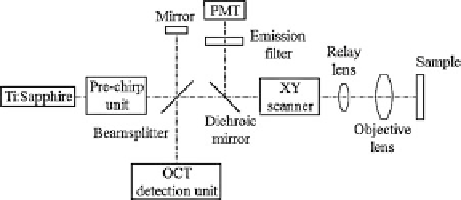
Fig. 9.5
Schematics of the
combined multiphoton and
optical coherence microscopy
system
Figure
9.5
is a multimodal imaging system using one light source, a Ti:sapphire
laser, that provides efficient multiphoton excitation and the broad bandwidth
required for high-resolution OCT. Pulse duration and bandwidth are selected
based on OCT resolution requirements and efficient fluorescence excitation. In the
illumination path, no excitation filter is needed because the excitation spectrum
is relatively monochromatic and is distinct from the emission spectrum. The
prechirp unit, consisting of prisms or a grating, precompensates the dispersion later
accumulated from the objective lens and from other optical components in the
beam delivery path. The beam is then split by a beamsplitter into two arms, the
sample arm and the OCT reference arm. The laser beam in the sample arm is raster
scanned by two galvanometer mirrors and relayed by the relay lens to the entrance
pupil of the objective lens. Then, the laser beam is focused onto the sample. The
fluorescence signal and backscattered signal are collected by the same objective
lens; the two signals are separated by the dichroic mirror after being descanned.
The fluorescence signal is directed to the photomultiplier tube (PMT) to construct a
multiphoton image, and the backscattered light is sent to the OCT detection path for
interference with the reference beam. The dichroic mirror in the detection path can
also be placed between the objective lens and the scan mirror for higher throughput.
Without being descanned, the emission light is not motionless at the detector plane
as the excitation beam is scanned across the sample. Therefore, the photosensitive
surface of the detector must be sufficiently large and the quantum efficiency should
be uniform. With simultaneous acquisition, perfect image registration is achieved,
providing spatiotemporal relationships between tissue structure and function.
Because both OCT and multiphoton imaging have high resolution, it is important
that the multimodal images be acquired from the same sampling volume. It is also
desirable that two modalities have matched transverse and axial resolutions. The
transverse and axial resolutions of MPM are determined by the NA of the objective
lens. The NA is usually large in order to focus the laser beam to a small volume for
high efficiency of nonlinear excitation. In OCT imaging, the transverse resolution is
similarly determined by the NA of the objective lens, but its axial resolution comes
from the coherence length of the light source, independent of the NA. Therefore, the
coherence length of the light source should be selected to match the axial resolutions
of multiphoton and OCT imaging. Tang et al. demonstrate that MPM and OCT
channels can be coregistered with lateral resolution of approximately 0:5 mand
axial resolution of approximately 1:5 m, utilizing an objective with 0.95 NA and
Search WWH ::

Custom Search