Biomedical Engineering Reference
In-Depth Information
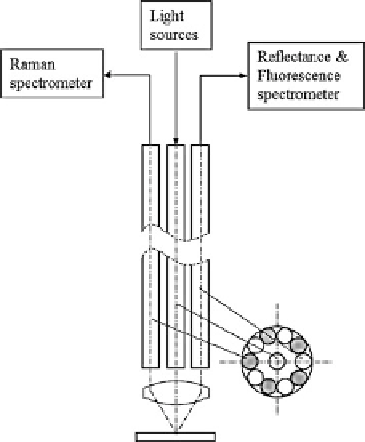
Fig. 9.4
Configuration of
multimodal spectroscopy. The
illumination light for each
modality is coupled into the
central fiber, and the signals
are captured by surrounding
fibers
to obtain promising results for discriminating breast cancer from benign breast
lesions [
23
].
9.2.5
Multimodal Multiphoton Imaging Systems
Both multiphoton microscopy (MPM) and OCT are capable of noninvasive, high-
resolution imaging in thick, scattering biological tissues. The contrast mechanisms
for these two techniques are intrinsically different in that the former is based on
the detection of fluorescence light through multiple photon absorption, whereas
the latter detects backscattered light through coherent gating. The combination
of OCT and multiphoton imaging offers complementary structural and functional
information about tissues and is a powerful imaging tool that has both the sensitivity
and specificity necessary to detect precancerous and cancerous lesions on both tissue
and cellular levels [
24
]. It also has the potential to identify a particular area of
interest through OCT and obtain high-resolution stack of multiphoton image of this
particular region.
Typically, OCT and multiphoton imaging require different light sources in order
to achieve optimal performance. Multiphoton imaging requires an ultrashort pulse
laser for efficient excitation, and OCT needs a light source with broad bandwidth
for high-resolution imaging. This is because multiphoton efficiency is proportional
to the inverse of the laser pulse duration and the axial resolution of OCT is inversely
proportional to the spectral bandwidth. While it is ideal to use different light source
for each modality, one light source is also possible for both modalities [
25
].
Search WWH ::

Custom Search