Biomedical Engineering Reference
In-Depth Information
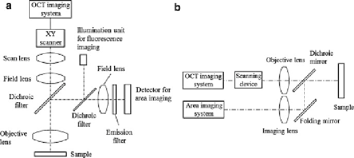
Fig. 9.2
Multimodal imaging system combining area imaging and scanning imaging modalities
through dichroic mirrors, (
a
) sharing the objective lens, and (
b
) without common component
not necessarily related to malignant transformation of tissue can be reduced with
OCT imaging. McNichols et al. developed a fluorescence-guided OCT endoscope
to detect oral cancer with potentially higher sensitivity and specificity [
5
]. This
multimodal imaging system allows rapid identification of suspected regions over
a large area using fluorescence imaging and obtains coregistered high-resolution
morphological features of the tissue using OCT. The abnormally increased fluores-
cence due to inflammatory reactions can be clearly differentiated from cancer by
OCT imaging; on the other hand, the atypical structure of a mature scar in OCT
image could be clarified with a fluorescence image. Pan et al. demonstrated that the
specificity of fluorescence detection of transitional cell carcinoma was significantly
enhanced by fluorescence-guided OCT (53 % versus 93 %), and the sensitivity
of fluorescence detection was improved by combining it with OCT (79 % versus
100 %) [
6
,
7
].
9.2.3
Multimodal Point Scanning Systems
Several imaging techniques discussed in Sect.
9.1
are point scanning techniques,
such as OCT, spectroscopy, and confocal imaging. Each of those point scanning
systems utilizes a different contrast mechanism and provides distinct information of
the tissue under investigation. However, each technique has its limitations.
OCT can provide detailed morphological information of the tissue in real time,
but it is sometimes difficult to interpret OCT images because contact of the device
with tissue can cause folds, stretching, or other localized tissue deformations.
Another issue with OCT imaging is that OCT images are simple maps of variations
in reflectivity and do not directly reveal the molecular composition of the tissue.
While spectroscopic techniques in general can extract tissue biochemical or
morphological information relevant to disease progression and diagnosis, each of
these techniques has its own limitations. For example, the spectra in fluorescence
Search WWH ::

Custom Search