Biomedical Engineering Reference
In-Depth Information
Tabl e 8. 3
Continuation of the table with examples of images obtained with various endomicro-
scopic modalities (contact imaging and optical coherence tomography)
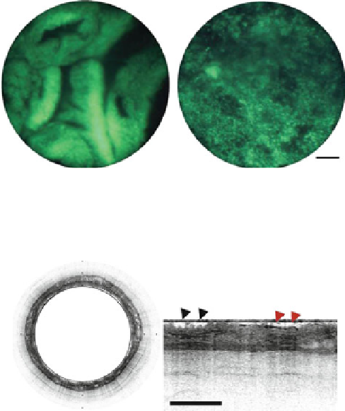
Contact imaging (fluorescence)
: Fiber bundle contact imaging with HRME system. Top: Barrett's
metaplasia and low-grade dysplasia and Bottom: esophageal adenocarcinoma. Scale bar
represents 100 m[
17
]
Optical coherence tomography
: Top: circular transverse cross-sectional Fourier-domain OCT in
vivo image of human Barrett's esophagus Bottom: expanded portion of the left figure pointing
surface irregularities (black arrowheads) and glands within the epithelium (red arrowheads).
Scale bar (right) and tick marks (left) represent 1 mm [
10
]
8.2.1.3
Imaging Depth
With confocal microscopy, optical sectioning can be achieved to depths rang-
ing from 200 to 400 m beneath the tissue surface, depending on the specific
wavelengths of excitation and emission, and the optical properties of the tissue
itself. Two-photon microscopy can reach greater depths due to the use of near-
infrared excitation wavelengths and the ability to collect all generated fluorescence
photons (no pinhole). Some recent experiments performed in the lab for 1,280-nm
wavelength allowed imaging depths of 0.9 mm [
18
]. Typically, however, in vivo
applications require lowering effective energy density and provide 200-500 m
imaging depths. Full-field contact imaging modes lack any intrinsic optical sec-
tioning ability, imaging only the most superficial tissue layers with good contrast.

Search WWH ::

Custom Search