Biomedical Engineering Reference
In-Depth Information
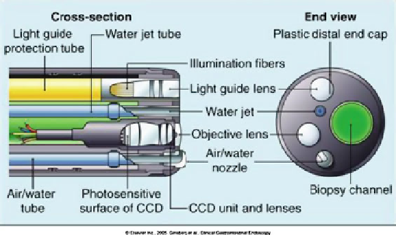
Fig. 8.1
Schematic cross section of the video endoscope tip [
1
]. The cross section demonstrates
illumination and detection tip components as well as biopsy channel and air/water nozzle
An ongoing challenge in endoscope design involves increasing functionality
and reducing endoscope diameter while including essential features such as an
accessory channel for biopsy collection and air and water channels for suction and
irrigation. Illumination channels are also required to deliver sufficient light intensity
to obtain a clear, bright image and are separated from the imaging path to eliminate
specular reflections and increase image contrast (Fig.
8.1
)[
1
]. While current state-
of-the-art endoscopes conceptually resemble their earlier counterparts, some novel
forms of endoscopy have been developed, received FDA approval, and entered
clinical practice. These novel techniques include for example capsule endoscopy
which uses a miniature camera encased in a “pill” which is swallowed by the
patient and transmits images as it passes through the gastrointestinal tract. Capsule
endoscopy offers the ability to evaluate sections which conventional upper or lower
GI endoscopes cannot access, such as the small intestine [
2
].
Endoscopic instruments are routinely used for medical diagnosis and treatment
of several organs, including the upper gastrointestinal tract (esophagus, stomach,
duodenum), lower gastrointestinal tract (colon, rectum), the airway, and lungs.
Depending on the intended application, the endoscope may range from around
3-13 mm in diameter and provide a 90-140
ı
field of view (FOV) with depth of field
ranging from 5-50 mm. In addition to conventional white light imaging, endoscopes
have also incorporated complementary modes including autofluorescence imaging
(AFI) [
3
] and narrow band imaging (NBI) [
4
-
6
]. AFI illuminates the tissue surface
with light at blue or UV wavelengths and collects only the longer wavelength
fluorescent light (green) emitted by the tissue. Changes in tissue structure due to
the development of cancer can alter the intensity of the fluorescent signal, assisting
the endoscopist in identifying abnormal areas. NBI illuminates the tissue with red,
green, and blue light within narrow wavelength bands instead of the full white light
spectrum. In NBI, the reflectance of tissue at these wavelength regions is altered by
the development of cancer and precancerous conditions. By digitally manipulating
Search WWH ::

Custom Search