Biomedical Engineering Reference
In-Depth Information
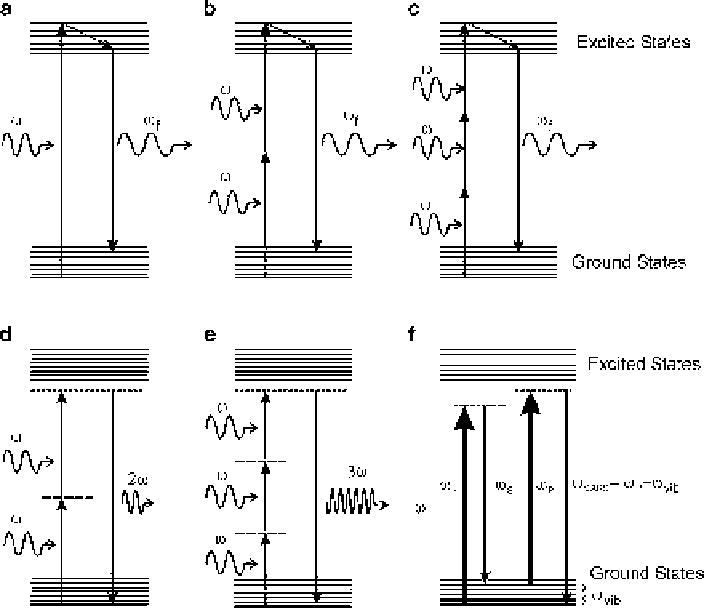
Fig. 7.1
Energy diagrams of various optical excitation methods that are used for imaging contrast
in biomedical field. The incoherent (nonresonant) modes of excitation resulting in fluorescence are
(
a
) 1-photon excitation, (
b
) 2-photon excitation, and (
c
) 3-photon excitation. The resonant coherent
modes of multiphoton excitation are (
d
) SHG, (
e
) THG, and (
f
) CARS resulting in a coherent signal
7.3
Multiphoton Imaging Modes
SHG, THG, and TPEF have become the most widely used contrast modes of
multiphoton imaging. The nonlinear contrast mechanism depends on the second-
and third-order nonlinearities as described by Eq.
7.1
, which can be produced by
the light-matter interaction at the focus of a high numerical aperture microscope
objective. As the nonlinear effects are proportional to the second or third power of
the fundamental light intensity, light only at a very tiny focal volume interacts with
the sample, eliminating the out-of-focus light, resulting in an inherent optical sec-
tioning. Selecting a nonlinear contrast method of choice, a multiphoton microscope
can be made by raster scanning a laser beam across the focal plane of a microscope
objective and measuring the signal intensity as a function of the focal spot position.
The 3-D images can be obtained by moving the sample or the objective along the
laser beam axis. A typical multiphoton imaging system is shown in Fig.
7.2
.
Search WWH ::

Custom Search