Biomedical Engineering Reference
In-Depth Information
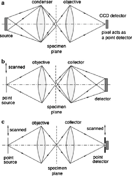
Fig. 6.7
The confocal microscope is compared with other forms of microscope: (
a
) a conventional
brightfield microscope with a CCD detector, (
b
) a scanning optical microscope, (
c
) a confocal
scanning optical microscope
there is also a small improvement in spatial resolution in confocal microscopy: we
return to this point later.
The confocal microscope is compared with other forms of microscope in
Fig.
6.7
. A conventional brightfield microscope with a CCD detector is illustrated
schematically in Fig.
6.7
a. This simplified diagram shows a microscope with critical
illumination: a large-area incoherent source is focused by a condenser lens on to
the specimen such that the entire field of the specimen is illuminated. Information
from each illuminated point in the specimen is simultaneously transmitted by the
objective lens to form an image, which is measured point by point by the pixels
of the detector. The important property to note is that it is the objective that is
responsible for forming the image, with the condenser playing only a secondary role
in determining the resolution of the system. In fact, the aperture of the condenser
Search WWH ::

Custom Search