Biomedical Engineering Reference
In-Depth Information
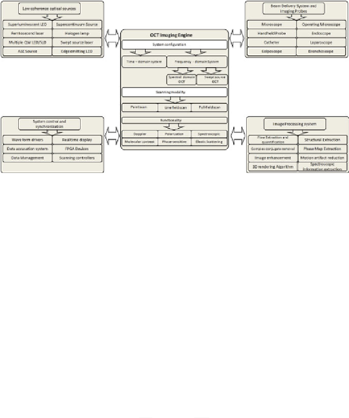
Fig. 5.4
A modular overview of OCT system with various peripheral components and schematic
of a typical first-generation free-space optics-based optical coherence tomography setup with
balanced heterodyne detection scheme
5.4
Practical Aspects of OCT System
5.4.1
Axial Resolution
Unlike the conventional optical microscopy, the apparent advantage of OCT is
that the axial resolution is completely decoupled from the lateral resolution. The
axial resolution of an OCT is an important specification of an OCT system, and in
many biomedical applications, high axial resolution is often required to distinguish
different cellular ultra-structures. As described in the preceding section, axial
resolution is defined as the full width at half maximum of the source coherence
length l
c
. For a source with a Gaussian spectral distribution, the axial resolution is
given by
0:44
2
2
2 ln 2
c
v
D
2 ln 2
z
D
;
(5.24)
where
z
and are the full width at half maximum of the autocorrelation function
and spectral width and
0
is the source center wavelength [
18
]. It can be seen that
broadband light sources are required to achieve high axial resolution, as the axial
resolution is inversely proportional to the spectral width of the light source. The
most popular light source currently used in OCT appears to be superluminescent
diode (SLD). A typical SLD has a bandwidth
D
20 nm centered at 830 nm,
which corresponds to a coherence length of
30 m, and since l
c
is defined for
a round trip, so this leads to a depth pixel size of 15 m in air, and in tissue, by
considering the refractive index of 1.4, gives
11 m. Modern sources for OCT
use Kerr-lens mode-locked lasers and photonic crystal fibers to achieve submicron
Search WWH ::

Custom Search