Biomedical Engineering Reference
In-Depth Information
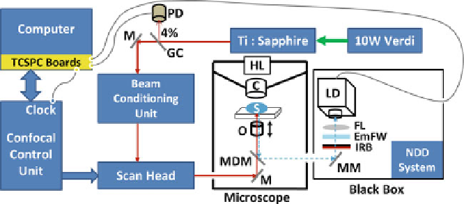
Fig. 3.6
The basic schematic of the TPE-TCSPC FLIM system. Details are described in
Sect.
3.4.2
. The excitation source is a 10 W Verdi pumped Ti:sapphire multiphoton (MP) laser,
which is coupled to the Bio-Rad Radiance 2,100 confocal/MP control unit for scanning the
specimen. The laser pulse reference is generated using a glass coverslip (GC) to reflect 4% of
the laser to a photodiode (PD) connected to the TCSPC device. Microscope: HL halogen lamp;
C condenser; S specimen; O objective lens; MDM movable dichroic mirror;
black box
:MM
movable mirror; IRB infrared light blocker filter; EmFW emission filter wheel; FL focusing
lens; LD lifetime detector; NDD system - the non-descanned detecting system for steady-state
multiphoton imaging; M - mirror
Our TPE-TCSPC FLIM system was first implemented in 2002 on a Bio-Rad
Radiance 2100 confocal/multiphoton (MP) microscope, using a TCSPC module
and a fast PMT detector purchased from Becker & Hickl (BH) [
148
]. The basic
schematic of the TPE-TCSPC FLIM system is illustrated in Fig.
3.6
. The Bio-
Rad system that is attached to a Nikon TE300 inverted epifluorescence microscope
and controlled using the LaserSharp 2000 software (
http://www.zeiss.com/micro
)
carries both single-photon (not pulsed) and MP lasers. The TCSPC FLIM system
is configured using the MP laser, which is a Coherent 10W Verdi pumped tunable
mode-locked ultrafast pulsed laser (Mira 900,
www.coherent.com
)
. The MP laser
has a repetition rate of 78 MHz and a pulsed width of less than 150 femtosec-
onds (fs) with a tunable range of wavelengths (700-1,000 nm). A laser spectrum
analyzer (Model E201, www.istcorp.com) is used to monitor the TPE wavelength,
and the power is measured at the specimen plane using a power meter (Model
SSIM-VIS & IR, www.coherent.com). The MP laser is coupled to the Bio-Rad
confocal/multiphoton control unit to scan the specimen via an XY raster scanning
mechanism using galvo mirrors.
A specific dichroic mirror (670UVDCLP) equipped on the microscope filter-cube
slider needs to be selected for fluorescence lifetime imaging, and this dichroic mirror
transmits the MP laser (670-1,000 nm) through the objective lens to excite the
specimen and reflects the emission light to the external (non-descanned) detectors
in a sealed black box. There are three non-descanned PMT detectors including the
lifetime detector and the other two PMT detectors that are used for the TPE steady-
state imaging. The emission light is routed by a movable mirror and then focused
Search WWH ::

Custom Search