Biomedical Engineering Reference
In-Depth Information
a
b
c
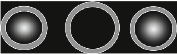
Fig. 8.7
Fabrication of a hemispherical electronic eye: (
a
) stretched hemispherical elastomer, (
b
)
array of detectors on the flattened elastomer, and (
c
) hemispherical electronic eye obtained after
releasing the elastomer
eye that has not only a three-dimensional refracting element but also a three-
dimensional arrangement of its detectors (
Ko et al. 2008
). This electronic eye is
illustrated in Fig.
8.7
. The hemispherical geometry, although suitable for a wide
field of view and low aberrations, is not compatible to the planar nanofabrication
techniques. Let us suppose that we want to fabricate a hemispherical eye camera
similar to the human eye, consisting of a number of pixels, the electronic part of each
pixel, referred to as silicon device, containing a photodiode and a blocking diode
monolithically integrated in a 500
500m
2
crystalline silicon and capped with a
micrometer thin polyimide layer. An ingenious trick is needed to place the silicon
devices manufactured using the usual wafer-scale two-dimensional technology on
a hemisphere: place first the two-dimensional array of Si devices on a flattened,
drumhead-like, radially stretched hemispherical elastomeric transfer element and
then release the elastomer to regain its hemispherical shape. The Si device array
adopts the same hemispherical shape due to van der Waals interactions with the
elastomer surface, but suffers no distortions if the interconnects between adjacent
Si devices are thin and narrow enough and have high elastic compressibility. In (
Ko
et al. 2008
), the interconnects were made from 50-m-wide, 360-m-long, and
3:150:3-nm-thick Cr:Au:Cr metal layers patterned on polyimide. These stages of
fabrication are illustrated in Fig.
8.7
. Afterward, the Si device array is transferred
onto a hemispherical glass substrate with the same radius of curvature and is
finally coated with a photocurable adhesive. Such a 16
16 pixels eye camera was
successfully fabricated and shown to function properly, and the poor image quality
due to the small number of pixels could be improved by collecting sequences of
images rotated in the azimuthal and polar directions by small increments and then
reconstructing the image (
Ko et al. 2008
); this strategy is also adopted in biological
systems.
In contrast to human and insect eyes, fish and octopus eyes use gradient index
biological structures to increase their focusing power and to correct spherical
aberrations. In particular, the octopus has no cornea so that it can see only with
the help of a spherical lens with an almost parabolic distribution of the refractive
index, obtained by a corresponding change of the water and protein concentrations.


Search WWH ::

Custom Search