Biomedical Engineering Reference
In-Depth Information
20.2 SLEEP BASICS
Sleep is a state of the brain, which is shared by most organisms from invertebrates to mammals.
The mechanisms underlying the transition from awake to sleep are now understood in some detail,
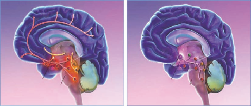
whereas the function of sleep still remains enigmatic. As illustrated in Figure 20.1, the transition
from wake to sleep is associated with a change in the activity of different neuronal pathways. Earlier
sleep was considered a resting state of the brain—an impression conveyed by the clinical signs of
sleep: reduced blood pressure, reduced heart rate, immobility, and reduced sensitivity toward exter-
nal stimuli. However, measurements of brain activity and imaging studies performed during sleep
have clearly shown that sleep is an active state of the brain. Sleep itself is not homogenous, and can
be divided into various states representing different patterns of electrical activity. The underlying
biology and complex interaction of various neurotransmitter systems in initiating, maintaining, and
shaping sleep are now beginning to be better understood, paving the way for more precise and effec-
tive pharmacological treatment of sleep disorders.
Like waking, sleep is an active state of the brain. During this heterogeneous and rapidly chang-
ing state several restorative functions take place, although the neural substrates of somatic and
cognitive restoration remain elusive.
Sleep is generally considered to consist of two substates, rapid eye movement (REM) and nonrapid
eye movement (NREM) sleep, which alternate to form a cycle lasting approximately 90 min (Figure
20.2). REM and NREM sleep can clearly be differentiated on the basis of a number of physiological
variables including muscle tone, electroencephalographic (EEG), and electromyelographic (EMG)
features, and the presence or absence of REMs. Distinct physiological roles for REM and NREM
stages have been proposed, but compelling empirical data are scarce.
Real sleep in a living brain is a continuous state without clear transitions. Therefore, a temporal
description of the waves and alterations in the amount of both frequencies and amplitudes should
most likely be based on an analysis of these waveforms. However, for historical reasons sleep stages
are described as either REM or NREM stages 1-4 using visual scoring criteria based, in part, on the
quantity and gross type of EEG waveforms per unit time. These are combined together graphically
into a hypnogram as shown in Figures 20.2 and 20.3. NREM stages 1 and 2 have been described as
Cerebral
cortex
Cerebral
cortex
Thalamus
Thalamus
Basal
forebrain
Basal
forebrain
PeF
Midbrain
Midbrain
vPAG
vPAG
LH
VLPO
LDT
PPT
LDT
PPT
BF
TMN
TMN
Raphe
Raphe
Cerebellum
Cerebellum
Hypothalamus
Hypothalamus
Pons
LC
LC
Pons
FIGURE 20.1
Neuronal pathways active during wake and sleep. Left: During wake, several arousal systems
are active. These include monoaminergic (noradrenalin and serotonin originating in LC, Raphe, and TMN)
and orexinergic (originating in LH) systems. Right: During sleep, the inhibitory GABA (VLPO) and melaton-
ergic systems (originating in the pineal gland and projecting to thalamus) take over and initiate and maintain
sleep and the transition between different sleep stages. PPT, pedunculopontine nuclei; LDT, laterodorsal teg-
mental nuclei; LC, locus coeruleus; TMN, tuberomammillary nucleus; vPAG, A10 cell group; LH, lateral
hypothalamus; BF, basal forebrain; VLPO, ventrolateral preoptic nucleus; PeF, perifornical neurons.