Biomedical Engineering Reference
In-Depth Information
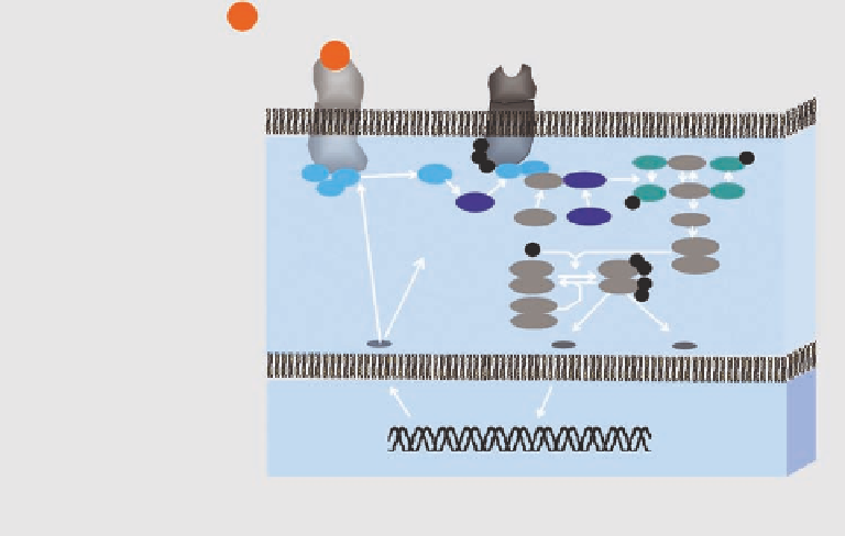
Ligand biodistribution
Extracellular
Receptor distribution and
occupancy
GPCR
NGFR
P
P
P
GTP
Ras
Ras
Raf
MEK1
GTP
Shc
Ga
Gb
Gb
Sos-1
Grb2
Gg
g
GDP
GDP
SRC
P
Grb2
Sos-1
Pathway activity
P
P
P
P
MEK2
ERK1
ERK1
ERK2
ERK2
PP2A
PTP1
Cytoplasm
System response:
morphological,
physiological, metabolic
cellular, and molecular readout
DNA
Nucleus
FIGURE 7.3
Imaging targets relevant for DDD. Currently available imaging techniques allow visualization
and quantii cation of the drug's mechanism of action. Labeling of the drug molecule itself (or of a competitive
receptor ligand) reveals information on its biodistribution and receptor interaction. The expression level of
a receptor can be visualized using specii c reporter ligands or following a reporter gene strategy. Activation
of the signaling cascade is visualized by targeting individual pathway molecules (e.g., caspases for studying
apoptosis) or by measuring protein-protein interaction (see text). Finally the result of the therapeutic interven-
tion such as morphological, physiological, metabolic, cellular, or molecular changes can be monitored.
reporter nuclide yields the sensitivity that is required to detect small amounts of the drug
ligand in tissue.
b.
Expression of the molecular target
: A critical step in early drug discovery is target valida-
tion, i.e., demonstration of the presence of a drug target in the tissue of interest. The com-
mon strategy to visualize and quantify the presence of a drug target, such as a membrane
receptor or an enzyme, uses target-specii c imaging probes, the vast majority using either
radionuclides or l uorescent dyes as reporter moiety. There are numerous examples of such
studies (Section 7.3.1). It is important to realize that unless one uses target-activatable
probes, it cannot be discriminated whether the signal observed arises from the reporter
fraction that is bound to its ta rget or just from free or unspecii cally bound molecules. Thus,
it is important to wait until the unbound probe is cleared from circulation. Alternatively,
reporter gene assays can be used to visualize target expression.
c.
Imaging pathway activities
: Two strategies can be pursued to study pathway activities,
either by monitoring critical molecules in the signal transduction cascade or by visual-
izing protein-protein interactions. The i rst approach uses the concept outlined in the
previous paragraph. For example, a reporter gene assay has been developed to visualize
the activity of caspases-3, a critical player in cellular apoptosis. Signal propagation relies
on protein-protein interactions. A number of assays have been developed to study these
key processes in cellular systems; some of them have been translated for applications
in intact animals such as the two-hybrid assay or the protein fragment complementation
assay. As an example, a split luciferase assay has been developed to study the interaction
of the two proteins FRB and FKBP12, which is induced by the administration of the
macrolide rapamycin.