Biomedical Engineering Reference
In-Depth Information
(a)
(b)
Submucosa
Epithelium
Elastic
fibres
Cartilage
Blood
vessel
Trachealis muscle
(c)
(d)
Intercartilaginous
zone
Reepithelial−
ization
Macrophage + fibroblast
infiltration
r
z
= +
h
/2
z
O
EPCs
MSCs
z
=−
h
/2
R
o
R
c
R
l
Epithelium
Cartilage
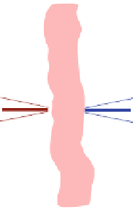
Fig. 1 Images of a a decellularised trachea, b the radial cross section of a healthy trachea, c an
axial cross-section of the trachea showing topical application of EPCs and MSCs, reepithelization
and infiltration of cells, and d a schematic diagram representing the cylindrical geometry of the
trachea, showing the position coordinates used in the mathematical model
to form a healthy functioning trachea, whereby the chondrocytes regenerated the
cartilage ring and the EPCs provided the lining of the airway (Fig.
1
b). The
therapy has been further developed
2
in the case of a young boy suffering from
congenital stenosis (abnormally narrow airway at birth) by seeding the donor
windpipe with autologously derived stem cells and EPCs in situ without the need
for seeding ex vivo. Improvements to the therapy are evolving rapidly, with the
most recent advance being the implantation of the first synthetic bio-engineered
trachea [P. Macchiarini, private communication].
2
BBC News, ''Windpipe transplant success in UK child'' Mar. 2010,
http://news.-
bbc.co.uk/1/hi/8576493.stm


Search WWH ::

Custom Search