Biomedical Engineering Reference
In-Depth Information
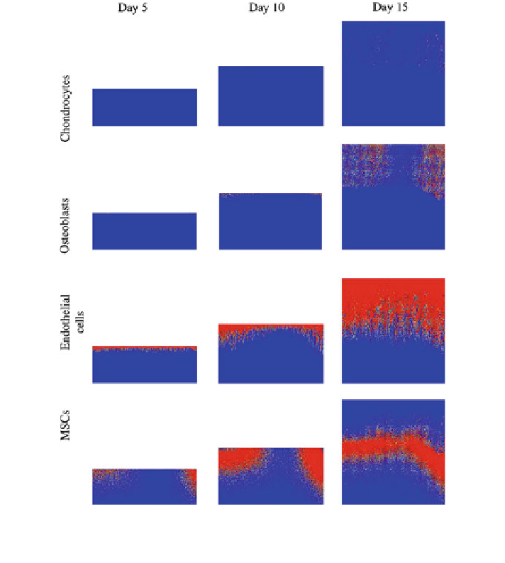
Fig. 6
Cell distribution prediction within the distracted gap during the first 15 days of the
process
walk model proposed by Pérez and Prendergast [
97
]. In addition the model
incorporates the vascular ingrowth in order to be able to capture the relationship
between bone formation and angiogenesis [
21
]. This capillary network is described
in terms of endothelial cell densities.
Figure
6
shows the evolution of four cell fates over the first 15 days of the
process of distraction: chondrocytes, osteoblasts, endothelial cells and mesen-
chymal stem cells (MSCs). It can be observed how this cell distribution varies
significantly. Initially, during the first days of the distraction procedure the gap is
gradually filled by MSCs that migrate from the marrow cavity, periosteum and
surrounding soft tissues to the interfragmentary gap. By day 10, osteoblasts could
be distinguished close to the old bone matrix. Osteoblasts differentiation is
assumed to be regulated by the vascular network. Figure
7
depicts the blood vessel
Search WWH ::

Custom Search