Biomedical Engineering Reference
In-Depth Information
(a)
(b)
(c)
(d)
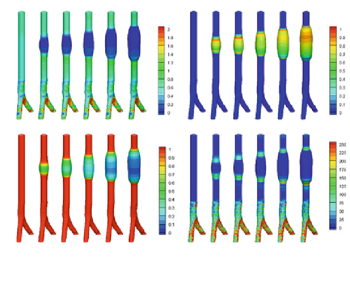
Evolution of wall shear stress s (a), degradation factor F
D
(b) elastin concentration m
E
Fig. 4
(c) and WSSG (d)att
¼
0
;
5
;
6
;
7
;
8
;
9 years
spatial distribution within the aneurysm, slightly larger magnitudes are observed
towards the proximal end. Also note that as with case (i), the axial strains in the parent
artery decrease as the aneurysm enlarges, i.e. the axial retraction of the ends of the
artery assists the axial expansion of the AAA. Figure
5
c illustrates the evolution of
the GL strains of positively wound collagen fibres in the media E
M
þ
:
At t
¼
0
;
the
collagen fabric is in material equilibrium, i.e. E
J
p
¼
E
AT
ð
J
¼
M
;
A; p
¼Þ
throughout the domain. Note that at t
¼
5
;
even though the geometry has changed
(and the elastin strains have increased) the collagen fabric is in homeostasis, i.e.
E
J
p
¼
E
AT
; the recruitment stretches and fibre concentration have evolved to restore
material equilibrium in the collagen fabric. As the elastin degrades and aneurysm
enlarges (t [ 5), the magnitude of the collagen strains increase; notice though that
the increases are small relative to the large deformation that is occurring—a con-
sequence of the remodelling of the reference configurations that the fibres are
recruited to load bearing. The average collagen concentration m
C
increases to
compensate for the loss of load borne by the elastin (Fig.
5
d).
Lastly, we consider the evolution of the cyclic stretch environment. Initially the
cyclic areal stretch is equal to 1.1 throughout the domain. At t
¼
5 slightly
elevated cyclic areal stretches (A
CS
¼
1
:
12) are present in the proximal and distal
necks of the aneurysm (see Fig.
5
e). As the elastin degrades and the collagen takes
over the load bearing, the cyclic areal stretch reduces to 1.04 within the aneurysm
Search WWH ::

Custom Search