Biomedical Engineering Reference
In-Depth Information
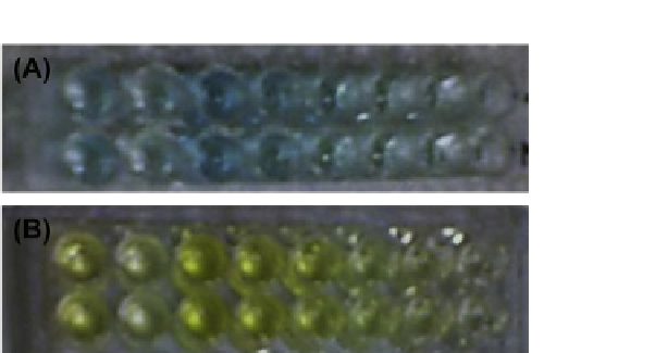
FIGURE 4.9
IOMNP-based capture of proteins from SK-BR3 breast cancer cells in an ELISA
using HRP enzyme label for detection. Signal after removing IOMNP ELISA (A) without stop solu-
tion and (B) with the stop solution.
Courtesy of Ocean NanoTech.
(For color version of this figure,
the reader is referred to the online version of this topic)
A quantitative detection of cells or proteins captured on a solid platform and
detected with fluorescence resulting from QDs attached to specific antibodies is
illustrated in
Figure 4.9
.
4.9 AN IOMNP-BASED ELISA FOR THE DETECTION
OF PROTEIN BIOMARKERS FOR CANCER
Aside from using the NMs such as AuNPs and QDs as signal generator, mag-
netic NMs such as the IOMNPs can also be used as the capture platform in
an ELISA. Similar to the magnetic beads, spherical IOMNPs have a high
surface area to volume ratio which allows for a better capture surface in het-
erogeneous assay. In addition, the IOMNPs do not interfere with the signal
detection because these can be easily removed from the detection solution by
magnetization.
4.9.1 A Protocol for IOMNP-Based ELISA Detection
of Breast Cancer Protein Biomarkers
The following protocol involves the development of a heterogeneous ELISA
using IOMNPs as the solid platform. The protocol involves IOMNPs that have
been previously used for magnetic NP cancer cell separation.
56
The IOMNPs
used in this protocol are 30 nm in diameter and larger ones can also be used for
faster magnetic separation.
(1)
Grow and harvest the SK-BR3 cells as in
Section 4.6.1
.
(2)
While growing the cells, conjugate anti-HER2 antibodies to 30 nM
IOMNP following the covalent conjugation protocol in Section 3.2.1.1
using IOMNP instead of the QDs.