Biomedical Engineering Reference
In-Depth Information
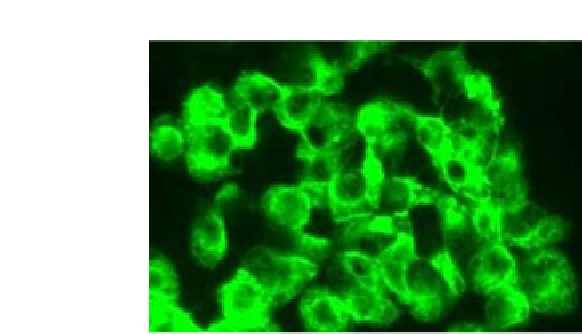
FIGURE 3.7
Adherent SK-BR3 cells on glass cover slip stained with QDs (530 nm).
Courtesy of
Ocean NanoTech, LLC. (For color version of this figure, the reader is referred to the online version
of this topic)
3.3.3.2 Minimizing PA of NMs on Biological Samples
To minimize nonspecific attachment of NMs on cells and other biological mate-
rials, washing and blocking buffers have been developed. These special buf-
fers contain blocking agents such as fish gelatin, bovine serum albumin (BSA),
casein, polyvinylpyrrolidone (PVP), ovalbumin, and other proteins that absorb
excess NMs during the staining process. These blockers are combined with sur-
factants and detergents such as sodium dodecyl sulfate, Tween 20, Triton X,
and others. Combined at proper ratios in buffer solutions such as 10 mM PBS,
pH 7.4 or 10 mM borate buffer, pH 7.4, the blocking agents and detergents help
eliminate nonspecifically adsorbed NMs.
195
The nonspecific adsorption of NMs could lead to clinical difficulties such as
failure of the medical application. Nonspecific adsorption of NMs can lead to
drug delivery to the wrong cell that can lead to a cascade of cellular processes
that may be triggered depending upon where the NMs are adsorbed causing
alteration to biological activity. In addition PA of NMs can lead to false posi-
tives when used for diagnostics.
Elimination of PA has been the focus of one study conducted by the group
of Aguilar.
195
In this study, they performed elimination of NMs' PA on cells in
vitro in the immunoassay detection of breast cancer cells using the SK-BR3 cell
line.
150,196
The studies involved the use of QDs that were functionalized with
-COOH on the surface. The QDs were used to conjugate Abs to form second-
ary Abs, 2°Ab-QD against the primary Abs that were used to label the surface
antigen HER2/neu, proteins found on the cell membrane that are overexpressed
in cancer cells. The complete assay consisted of the SK-BR3 + anti-HER2/neu
+ 2°Ab-QD while the incomplete assay did not contain the specific cell mem-
brane label, anti-HER2/neu. The process used to eliminate PA is as follows:
(1)
Grow the cells in a culture flask following the manufacturer's recommen-
dations.