Biomedical Engineering Reference
In-Depth Information
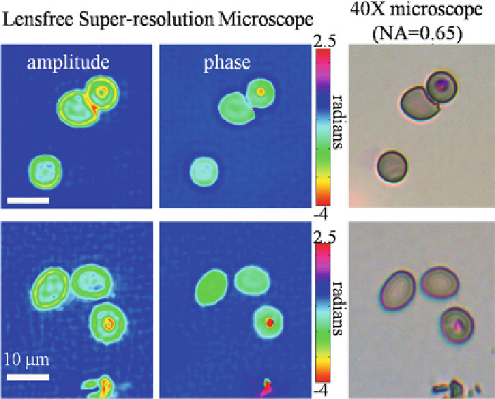
Fig. 4.11
Images of red blood cells infected with malaria parasites in a thin blood smear, obtained
using the field-portable lensfree super-resolution microscope (see Fig.
4.9
). The parasites within
the cells are visible in the amplitude and phase images. A bright-field microscope image is also
provided for comparison, in which the parasites have a different color due to staining
corresponding microscope image are compared to the super-resolved hologram
obtained from the PSR algorithm. The low-resolution hologram exhibits aliasing
due to undersampling, which is evident from the curvature of outer fringes. After
combining the multiple frames using PSR, this spatial aliasing is resolved and the
curvature of the holographic fringes changes accordingly. These resolved fringes
translate to a larger effective numerical aperture, which leads to better resolved
microscope images after the phase-retrieval processing described in earlier sections.
The object that was imaged is a “UCLA” pattern etched in glass using focused ion
beam (FIB) milling; therefore, it is nearly a phase-only object. The width of the
letters and spacing between them in this pattern are on the order of 1m, and they
are visibly resolved in the super-resolution microscope image.
The PSR holographic on-chip microscope is designed to be lightweight, robust,
and cost-effective, with global health-related applications in mind, such as disease
diagnostics or water quality monitoring. As an initial demonstration of the capa-
bilities of this microscope in tackling such issues, this field-portable device was
used to image red blood cells infected with malaria parasites (
P. falciparum
)ina
standard thin blood smear. Figure
4.11
shows the PSR microscope images of healthy