Biomedical Engineering Reference
In-Depth Information
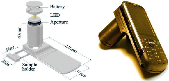
Fig. 4.6
Schematic illustration (
left
) and a photograph (
right
) of the lensfree holographic cell
phone microscope are shown. Weighing
38 g, this mechanical attachment converts a regular cell
phone with an existing CMOS sensor chip to a lensfree on-chip microscope
platforms running on cell phones [
25
,
35
-
38
]. For instance, a mobile phone-
mounted light microscope has been demonstrated that is capable of bright-field and
fluorescence imaging to identify
Plasmodium falciparum
-infected and sickle red
blood cells as well as
Mycobacterium tuberculosis
-infected sputum samples [
25
].
Alongthesamelines,wehavealsodeveloped a compact and cost-effective
microscope integrated on a cell phone [
11
] that does not utilize lenses and other
bulky optical components. This telemedicine microscope, shown in Fig.
4.6
, is based
on the partially coherent lensfree digital in-line holography technique introduced
in the previous section and inherits its advantages such as large field of view
(e.g.,
24 mm
2
), architectural simplicity, and mechanical robustness. This mobile
platform utilizes a lightweight add-on unit that attaches to the cell phone to convert
it to a telemedicine microscope. The add-on unit simply consists of a battery-
operated LED (center wavelength at 587 nm) that is butt-coupled to a large pinhole
(
100m diameter), a hollow tube for light propagation and a sample-loading tray
to mount the objects on top of the built-in digital sensor of the cell-phone camera
unit (5 MP,
24 mm
2
active area), whose lens is physically removed. The objects
placed on the sensor with <2 mm distance to its active area are then illuminated
by the installed LED to record digital in-line holograms of the objects using the
color (i.e., RGB) sensor chip of the cell phone. Nevertheless, the sensors that are
employed in cell-phone cameras comprise color filters tiled in a Bayer pattern,
which are optimized for color photography. This renders the cell-phone sensors
nonideal for holographic microscopy, where a quasi-monochromatic light source
(e.g., an LED) is employed for illumination, as these color filters lead to nonuniform
pixel response partially distorting the holographic information. To minimize this
distortion due to color filters, we utilized an additional digital correction step in
our holographic reconstruction algorithm, summarized in Fig.
4.7
, whose aim is to