Biomedical Engineering Reference
In-Depth Information
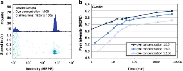
Fig. 3.6
Left:
Intensity histogram and speed profile for
Giardia lamblia
cysts measured with pro-
totype instrument shown in fig.
3.5
Right:
Incubation study for
Giardia lamblia.
Our experiments
showed that even for an analyte/reagent ratio of 100:1 a staining time of
2 min is sufficient
for reliable detection. We used a very simple sample preparation: mixing, incubation, no wash.
On-chip sample prep seems straight forward for this test
3.4
Multiplexed Flow Assay
For advanced diagnostics, multiple biomarkers within a complex biological sample
need to be simultaneously detected and quantified (proteins, cells, DNA fragments,
(bio)molecules, etc.). For quantification of cellular markers, for example, CD4 cells,
flow cytometry is an obvious choice. The size of cells is well compatible with the
flow cytometer technology. Thousands of staining assays provide the sensitivity and
specificity for intra- and cell surface markers that can be detected, enumerated, and
quantified in flow cytometers. The detection of smaller individual bioparticles, for
example, viruses, is less common [
45
] because fewer specific stains are available,
fluorescent brightness and scatter signals are generally lower, and the small size
of objects leads to inaccurate detection when multiple particles are present in
the detection area. Especially the latter failure mechanism prevents specific direct
quantification of biomolecules (e.g., proteins, DNA fragments) in solution.
In order to detect and quantify incorporated dyes of (bio-)molecules simultane-
ously, various detection schemes have been developed - enzyme-linked immunosor-
bent assay (ELISA) [
46
,
47
], DNA microchip [
48
,
49
], multiplexed SPR [
50
,
51
], etc.
The common characteristic of these techniques is the fact that different detection
reagents are spaced in close proximity and the detection scheme takes position-
resolved measurements. Different positions therefore identify different analytes. For
any of these techniques, the detection positions are located on a surface, making the
lateral diffusion lengths and times of analytes the relevant ones for the measurement.
For flow cytometer applications, multiplexed (fluorescent) particle-based assay
have been developed and commercialized. These assays consist of different types
of beads which can be distinguished by size (e.g., Assay Designs), emission