Biomedical Engineering Reference
In-Depth Information
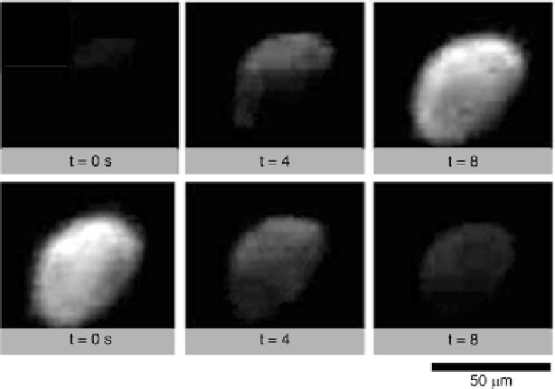
Fig. 2.11
Vesicles were electroporated using a hybrid integrated circuit/microfluidic device. At
t
0 sinthe
upper left corner
, a vesicle surrounded by fluid containing fluorescein, a fluorescent
molecule, was electroporated. The molecule entered the vesicle, making it fluoresce at t
D
D
8 s. At
time t
0 sinthe
lower right corner
, a fluorescent vesicle was electroporated. The fluorescein
diffused into the surrounding fluid
D
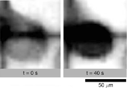
Fig. 2.12
A cell is
electroporated using the
hybrid integrated
circuit/microfluidic chip. The
cell membrane is normally
impermeable to trypan blue, a
dye commonly used to stain
cells, but after
electroporation, it enters and
darkens the cell
Cells can also be electroporated to insert contents into the cell and to release
its contents, as seen in Fig.
2.12
. This is a critical function enabling biological
vectors and functionalized particles to be introduced into the cell and to analyze
the cell contents [
22
-
24
]. Cell electroporation is demonstrated by placing a yeast
cell on the hybrid integrated circuit/microfluidic chip in a solution containing
trypan blue, a dye commonly used for staining biological samples [
10
,
12
]. The
cell membrane is impermeable to trypan blue. The cell was trapped above a