Biomedical Engineering Reference
In-Depth Information
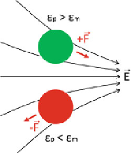
Fig. 2.2
An illustration of
positive and negative
dielectrophoresis (DEP). The
top
object is undergoing
positive
DEP, being pulled
toward
the electric field
maxima. Its dielectric
constant is greater than that of
the surrounding medium. The
bottom
object is being pushed
away
from the maximum by
negative
DEP. Its dielectric
constant is less than that of
the surrounding medium
Fig. 2.3
A plot of three frequency regimes for vesicles. In the 1-Hz-1-kHz range, electroporation
and electrofusion dominate. In the 1-kHz-10-MHz range, dielectrophoresis (
DEP
) dominates.
Above 1 GHz, microwave heating dominates [
10
]
electric field, and "
p
is the particle's permittivity. The strength and sign up the DEP
force depends heavily on the frequency of the electric field, which is embedded in
the complex permittivities in the Clausius-Mossotti factor. DEP for cells floating
in water is optimal at a frequency of approximately 1 MHz. This is illustrated in
Fig.
2.3
with the curve labeled “DEP.”
As shown in Fig.
2.3
, different functions can be performed on cells and vesicles
at various electric field frequencies. At low frequencies, cells can be porated and
fused. The 1-Hz-1-kHz range destabilizes vesicles by creating a potential across
their membranes. A large enough potential causes dielectric breakdown of the