Biomedical Engineering Reference
In-Depth Information
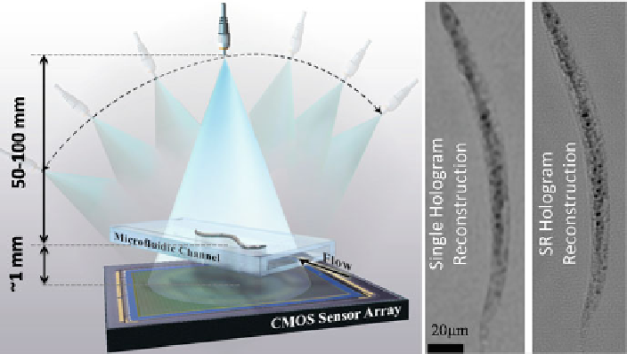
Fig. 4.17
A schematic of holographic opto-fluidic microscopy (HOM), which can provide super-
resolution (SR) holograms of flowing objects within the micro-channel at any illumination angle
(
left
). For this end, multiple lensless holograms are captured at a given illumination angle as the
object flows through the microfluidic channel. To the
right
, the images of a
Caenorhabditis elegans
worm with vertical illumination are shown, where the multi-frame PSR result shows noticeably
enhanced resolution when compared to the single hologram microscope image. In this case, 15
shifted holograms were used as input. Using different viewing angles within an angular range of
˙
50
ı
, the same platform can also serve as an opto-fluidic tomographic microscope as illustrated
in Fig.
4.18
a
C. elegans
worm is shown. If a single hologram is used to recover a microscopic
image of the worm (ignoring the flow), the resolution of the reconstructed image is
constrained by the physical pixel size of the CMOS sensor, as we discussed earlier.
However, when the flow is utilized to capture multiple frames for PSR processing,
the resulting reconstructed microscopic image is sharper and reveals more details
and better contrast due to the smaller effective pixel size after PSR processing (see
Fig.
4.17
). In this case, 15 shifted consecutive holograms were used for PSR, which
took 3 s of acquisition time at 5 frames per second, with the object flowing at a
speed of
1m per second. A higher frame rate sensor would allow a shorter total
acquisition time and faster flow of the samples within the biochip.
Owing to the almost alignment-free, robust, and simple nature of our illumination
scheme, the same holographic optofluidic microscope (HOM) can be conveniently
converted to an optofluidic tomography platform as shown in Fig.
4.17
[
21
]. By
illuminating the objects from different angles as they flow through the microchannel,
it is possible to record multiple projection holograms for each angle and then
synthesize SR holograms of the objects, as described above for a
C. elegans
worm that is electrokinetically driven through a microchannel. Using the procedure
described in Sect.
4.6
, these SR holograms can then be used to compute tomograms
of the flowing objects. As shown in Fig.
4.18
, optofluidic tomography can achieve