Biomedical Engineering Reference
In-Depth Information
Figure 13.1
M
2
A
TM
Capsule Endoscopy
Given
TM
Diagnostic System, including a wire-
less capsule endoscope (lower left), sensors for receiving data (middle), a portable
data recorder(lower right), and a workstation (upper) for viewing the images.
(From
www.givenimaing.com
). For color reference, see page 221.
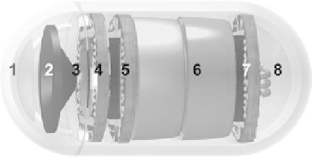
Figure 13.2
Schematic drawing of components inside the capsule endoscope. 1-optical
dome, 2-lens holder, 3-lens, 4-illuminating LEDs, 5-CMOS image sensor, 6-battery, 7-ASIC
transmitter, 8-antenna. For color reference, see page 221.
Prior to undergoing wireless capsule endoscopy, the patient is required to
have a 10-hour fast. The procedure begins after the capsule is ingested with a
sip of water. Then the capsule moves through the GI tract passively by natural
peristalsis and captures 2 images per second. During the procedure, the sensor
array is attached to the patient's skin in the abdominal region, and the other end
is connected to the data recorder, which is worn by the patient on a belt around
the waist. Images transmitted by the capsule at 433.10MHz through the built-in
antenna are picked up by the sensor array and then sent to the recorder.
The procedure usually takes about 8 hours, during which more than 55, 000
images are acquired. At the conclusion of the examination, the patient returns the
belt and the recording unit to the physician. Then the physician can download,
view, edit and archive data images on the workstation, which is a computer