Biomedical Engineering Reference
In-Depth Information
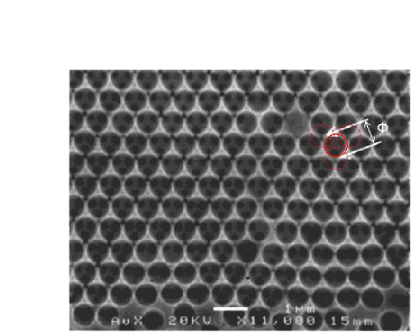
conditions. This is clearly shown in the following figure if more than
one layer of PS is used in templates.
SEM images of regions of macroporous gold films grown with
a thickness gradient by electrochemical deposition through templates
assembled from either 750-nm-diameter polystyrene spheres [204]. The
electrodeposition was performed at a potential of
Figure 1.18
. Φ is the
diameter of the sphere on top layer. The three dotted circle lines represent
the spheres beneath. Reproduced by kind permission from the publisher.
-
0.90 V
SCE
The SEM image shows that the spherical voids left in the gold
films after the removal of the PS spheres are arranged in well-
ordered, single domain, close-packed structures. Fig. 1.
18
gives an
image for a macroporous gold film prepared through a template of
750-nm-diameter spheres in a region where more than one layer of
PS is self-assembled. Within each hemispherical void in the top layer
there are again three smaller dark circles (diameter ca.100 nm).
These correspond to the interconnections to the three spherical
voids in the layer below (marked as dotted circles in Fig. 1.
18
) that
are left around the regions where the original polystyrene spheres
in the two layers were in contact. Semiconductors such as PbO
can
2


Search WWH ::

Custom Search